Perfusion, Circulation and Sleep
How to Use This Guide
The eight figure questions — a fifth of the paper — all come from Part 3: Where Each Valve Is Heard — APETM, Finding the Spaces by the Ribs (the rib-cage find-it game) and Where the Valve Is, and Where You Listen.
20 minutes
The night before
- Rapid Review — the marked items, then the traps.
- One clean round of the rib-cage game in Finding the Spaces by the Ribs.
An hour
The high-yield hour
- The Path in One Line, then Part 3 from APETM on — both find-it games, the extra sounds, rubs and bruits.
- Part 8, Rest and Sleep — six of the forty questions — and its self-test.
- Rapid Review.
The full guide, with the audio
Over several sittings
- Parts 1 to 4 — the heart: its path, its sounds, what backs up where. They are the Quiz 3 guide’s Parts 1 to 4, so skim what you already know cold.
- Part 5 — the cardiac assessment and its tests.
- Parts 6 and 7 — the peripheral vascular assessment and its disorders.
- Part 8, then Appendix A (the week’s terms), Rapid Review and the Summary Recap.
Most parts end with a self-test. On a phone, wide figures pan sideways, wide tables turn into one card per row, and every self-test answer stays hidden until you tap Show answer.
The Path in One Line
Say it out loud until it runs on its own. A fill-in-the-blank version hands you the venae cavae; after that it is one chain, because blood only goes one way through a healthy heart. Learn it as two halves of seven: right side to the lungs, left side to the body.
| # | Stop | Kind | What happens there |
|---|---|---|---|
| 1 | Superior & inferior vena cava | Great veins | Deoxygenated blood returns from the body — the superior from the head and arms, the inferior from everything below the heart. Fill-in-the-blank versions usually give you this one. |
| 2 | Right atrium | Chamber | The receiving room on the right. Both cavae empty into it. |
| 3 | Tricuspid valve | Valve (AV) | The door from the right atrium down into the right ventricle. Three cusps. |
| 4 | Right ventricle | Chamber | Pumps the blood out to the lungs. A short, low-pressure trip, so a thinner wall than the left. |
| 5 | Pulmonic valve (also: pulmonary semilunar valve) | Valve (semilunar) | The exit door of the right ventricle. |
| 6 | Pulmonary arteries (right & left) | Great arteries | Carry the blood to the lungs. The only arteries in the body that carry deoxygenated blood. |
| 7 | Lungs | Gas exchange | Carbon dioxide out, oxygen in, across the pulmonary capillaries. The colour changes here, and only here. |
| 8 | Pulmonary veins (four — two from each lung) | Great veins | Bring the fresh blood back to the heart. The only veins in the body that carry oxygenated blood. |
| 9 | Left atrium | Chamber | The receiving room on the left. |
| 10 | Mitral valve (also: bicuspid valve) | Valve (AV) | The door from the left atrium down into the left ventricle. Two cusps. |
| 11 | Left ventricle | Chamber | The strongest chamber, with the thickest wall — it pumps to the whole body against the highest pressure. |
| 12 | Aortic valve (also: aortic semilunar valve) | Valve (semilunar) | The exit door of the left ventricle. |
| 13 | Aorta | Great artery | The largest artery in the body. Arches over the top of the heart and carries blood to everything. |
| 14 | The body | Gas exchange | The tissues take the oxygen, the blood turns deoxygenated, and the veins bring it back to stop 1. |
“Venae cavae, right atrium, tricuspid, right ventricle, pulmonic, pulmonary arteries, lungs.”
“Pulmonary veins, left atrium, mitral, left ventricle, aortic, aorta, body.”
The Loop
The fourteen stops bent into a circle: start at stop 1, bottom left, up the blue side, across the lungs, down the red side. The heart is two pumps side by side, in one loop, so they move the same volume with every beat; the left is stronger because it pushes against higher pressure, not because it moves more. (Standard physiology; the deck defines stroke volume and cardiac output but does not compare the two sides.) And the colour changes in exactly two places — red in the lungs, blue in the body — never inside the heart, because the septum keeps the sides apart.
Two games. Trace it blanks every label and number and gives you a word bank: pick the stops in order, starting after the venae cavae, and each one fills in as you get it — exactly what a fill-in-the-blank question asks. What comes next? lights one stop, hides the rest, and asks you to click the one after it.
Drag sideways to see the whole figure →
Inside the Heart
The lab worksheet, and very likely any test figure, uses an anatomical outline, so find the same stops on a real shape.
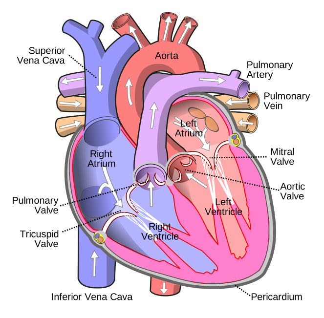
| Look for | How to recognise it |
|---|---|
| The two cavae | The vertical vessel on the left of the page, entering the right atrium from above (superior) and below (inferior). |
| The aorta | The arch over the top, three branches rising to the head and arms. It leaves the left ventricle and curves up and over to the patient’s left, so its root is mid-heart. |
| The pulmonary trunk | In front of the aorta, leaving the right ventricle and splitting into right and left pulmonary arteries; the right one passes behind the aorta. |
| The pulmonary veins | Short stubs entering the left atrium from the sides — usually two each side, four in all. |
| The ventricles | The left has a much thicker wall — the easiest tell on an unlabelled drawing. |
| The apex | The tip, down and to the patient’s left, formed by the left ventricle. Not a stop. |
The Word Bank
The only word bank the course has shown you is on the heart lab worksheet, where you labelled the heart and traced the flow in red and blue. Its terms, sorted by whether each is a stop:
| Term | On the path? | Where it goes |
|---|---|---|
| Superior Vena Cava | Yes | Stop 1 — usually given |
| Inferior Vena Cava | Yes | Stop 1 — usually given |
| Right Atrium | Yes | Stop 2 |
| Tricuspid Valve | Yes | Stop 3 |
| Right Ventricle | Yes | Stop 4 |
| Right Pulmonary Artery | Yes | Stop 6, to the right lung |
| Left Pulmonary Artery | Yes | Stop 6, to the left lung |
| Right Pulmonary Vein | Yes | Stop 8, from the right lung |
| Left Pulmonary Veins | Yes | Stop 8, from the left lung |
| Left Atrium | Yes | Stop 9 |
| Mitral Valve | Yes | Stop 10 |
| Left Ventricle | Yes | Stop 11 |
| Aorta | Yes | Stop 13 |
| Apex | No | A landmark — the tip of the left ventricle. Labelled on the diagram, never filled into a flow blank. |
The documents disagree on ten blanks or eleven (see the endnote); you cannot predict the subset, so learn the whole chain. Part 2 drills every version.
One Name, Several Labels
A blank that wants “mitral” may sit beside a diagram that says “bicuspid”. A change of label is not a change of answer.
| Course name | Also called | Why |
|---|---|---|
| Tricuspid valve | Right AV (atrioventricular) valve | Right atrium to right ventricle; three cusps. |
| Mitral valve | Bicuspid valve · left AV valve | Two cusps, like a bishop’s mitre. Mitral and bicuspid are the same valve. |
| Pulmonic valve | Pulmonary (semilunar) valve | The deck says pulmonic, its valve figure pulmonary. Both are right. |
| Aortic valve | Aortic semilunar valve | Half-moon cusps, like the pulmonic. |
| Pulmonary arteries | Pulmonary trunk, then right & left pulmonary arteries | A blank may want the trunk, the singular or the plural. |
| Vena cava | Superior & inferior · plural venae cavae | Two vessels, one destination — the right atrium. |
The atrioventricular-and-semilunar terminology is standard anatomy rather than something the cardiac deck spells out; it is how the four valves sort into the two heart sounds in Part 3.
The Quiz, Rehearsed
The blood-flow question as the Quiz 3 blueprint described it: venae cavae given, the rest blank, a word bank with Apex left in to reject. Type into each blank, or click a blank and then a word.
Three versions, because the documents disagree on the count. Eleven is right atrium to aorta with all four valves (the peripheral-vascular prep sheet’s count). Thirteen adds the lungs and the body. Nine drops the two semilunar valves, as the worksheet’s bank does. When you can do all three with the bank hidden, you are ready.
Two Kinds of Valve
The four valves come in two pairs, and the pairing is the key to the heart sounds. Each lets blood through one way only — the Think Conceptually handout’s “Normal Function of the Heart Valves (open and closing)”. (Standard physiology: valves have no muscle; they open and shut passively, on pressure.)
| Pair | Valves | Sits between | Shuts when… and makes |
|---|---|---|---|
| Atrioventricular (AV) | Tricuspid (right) Mitral (left) | Each atrium and the ventricle below it | The ventricles start to squeeze — start of systole. Together they make S1, “lub”. |
| Semilunar | Pulmonic (right) Aortic (left) | Each ventricle and its great artery — pulmonary trunk, aorta | The ventricles stop squeezing and relax — start of diastole. Together they make S2, “dub”. |
(Standard anatomy, not in the deck: the AV valves are held by chordae tendineae on papillary muscles, which stop the flaps blowing back; semilunar cusps simply fill and seal.)
Lub and Dub
The deck: systole is “myocardial contraction”, diastole the “filling phase”; Jeopardy: the phase “related to relaxing of the heart muscle” is diastole. The two sounds are the borders between the phases.
| Sound | Valves closing | When | Loudest at |
|---|---|---|---|
| S1 “lub” | Mitral and tricuspid — the AV valves | Start of systole | The apex — tricuspid and mitral areas |
| S2 “dub” | Aortic and pulmonic — the semilunar valves | Start of diastole — “marking the end of systole” | The base — aortic and pulmonic areas |
Drag sideways to see the whole figure →
The clips are played two octaves higher than life so a phone or laptop speaker can carry them; with headphones, open “Original pitch” under each player to hear the true sound a stethoscope gives.
Original pitch, for headphones
Where Each Valve Is Heard — APETM
The largest block on the Exam 4 blueprint: 8 questions with skeletal figures and locating heart sounds. The five listening posts, top to bottom, spell APETM.
Heart lab prep sheet: “APETM designation… understand the landmarks, where S1S2 heard loudest and why (will be tested).”
Exam 4 blueprint: “Must be able to name and document the locations (on a skeletal figure) to auscultate each of the heart sounds. (APETM, full name, and where)”; “Identify the locations of prominent S1 and S2 on the same skeletal figure”; “Auscultating apical pulse and location on a skeletal figure”.
So for each site: the letter, the full name, the intercostal space and border, and which sound is louder there.
| Full name | Where | Louder | Why | |
|---|---|---|---|---|
| A | Aortic | 2nd ICS, right sternal border — the only site on the patient’s right | S2 > S1 | Base. S2 is the aortic and pulmonic valves closing, and the sound carries up, with the flow, into the great arteries. |
| P | Pulmonic | 2nd ICS, left sternal border | S2 > S1 | Base. Same reason, on the pulmonary trunk’s side. |
| E | Erb’s point | 3rd ICS, left sternal border | S1 = S2 | Halfway between base and apex — the deck’s “point at which valves are usually equally audible”. |
| T | Tricuspid | 4th ICS, lower left sternal border | S1 > S2 | Apex. S1 is the tricuspid and mitral closing, and the sound carries down, with the flow, into the ventricles. |
| M | Mitral | 5th ICS, left midclavicular line — the apex, the PMI, the apical pulse | S1 > S2 | Apex. Same reason, at the tip of the left ventricle. |
The lab manual words the tricuspid row “S1 > or = S2”; use the deck’s five lines (see the endnote).
This works like the skeletal figure on Exam 4. Start hides every letter and label and names a site — click where you would put your stethoscope. Then say its intercostal space and which sound is louder there before you look.
Drag sideways to see the whole figure →
Lab manual: normally 1–2 cm and brief, often not palpable in adults, displaced laterally and left in late pregnancy. Slide-16 notes say “4th–5th ICS”; answer fifth, though “4th–5th” is not wrong (endnote).
The technique, in the deck’s order
- Rate and rhythm first, in one area — “usually apex or Erb’s point”. Regular: count 30 seconds and multiply by 2. Irregular: count a full minute.
- Then each area: S1, S2 and any extra sounds.
- Erb’s point is the alternative apical-pulse site — the deck’s notes: “Often easier than Apex for large breasted women.”
- Pulse deficit = apical − radial (Jeopardy), for an irregular pulse: 96 at the apex, 80 at the wrist → 16 beats too weak to make a pulse. (Standard: two nurses, same full minute.)
- Draping: displace breast tissue laterally; expose only the part of the chest being assessed.
Bell or Diaphragm
Equipment: a stethoscope and a watch with a second hand. Lab manual: “Listen with bell (light pressure) and diaphragm (heavy pressure) of stethoscope at all sites.”
| Side | How to hold it | Best for |
|---|---|---|
| Diaphragm (the wide, flat side) | Firm pressure | High-pitched sounds: S1 and S2, and most murmurs |
| Bell (the small, cupped side) | Light pressure — just enough to seal | Low-pitched sounds: S3 and S4, the rumble of mitral stenosis, and bruits over an artery |
Finding the Spaces by the Ribs
On a drawing, the only thing you can count is ribs, so learn each site as a rib address.
Rib addresses: aortic and pulmonic between ribs 2 and 3, Erb’s 3–4, tricuspid 4–5, mitral 5–6 at the midclavicular line.
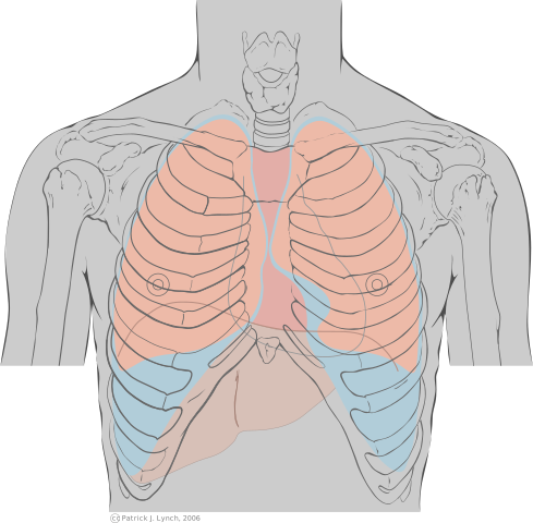
Press Start. The unmarked copy has no dots: count down from the sternal angle and click where each site is. Some questions ask by name, some by rib address, some by which sound is louder.
Where the Valve Is, and Where You Listen
Two maps of the same four valves. Anatomy: all four bunched behind the sternum, from the pulmonic (highest) to the tricuspid (lowest), where their sounds overlap (standard anatomy). Auscultation — APETM — listens downstream instead.
Switch between the two maps, then show both to see how each valve’s sound travels to its post.
Drag sideways to see the whole figure →
| Valve | Where it sits | Where you listen | Why there |
|---|---|---|---|
| Aortic | Behind the left half of the sternum, about the 3rd ICS | 2nd right ICS, RSB | The aorta rises up and to the right before it arches — so a left-sided valve is heard on the right. |
| Pulmonic | At the left 3rd costal cartilage — highest | 2nd left ICS, LSB | The pulmonary trunk runs up and to the left. |
| Tricuspid | Behind the right half, about the 4th ICS — lowest | 4th left ICS, LLSB | Into the right ventricle, behind the lower sternum. |
| Mitral | Behind the left half, about the 4th costal cartilage | 5th left ICS, MCL — the apex | Into the left ventricle, which points down and out. |
| Erb’s point | No valve of its own | 3rd left ICS, LSB | Between base and apex: S1 and S2 about equal. |
Extra Sounds
Anything beyond two syllables is an extra sound; the sound strip under Lub and Dub marks where each falls.
S3 and S4 — the gallops
Both are quiet, low thuds in diastole from blood hitting a ventricle that cannot take it. The deck: S3 is “rarely normal in older adults”; S4 comes “late in diastole” from “abnormal resistance to ventricular filling”.
The deck lists children among those in whom S4 can be normal; standard teaching says it is S3 that is normal in children (see the endnote).
| S3 | S4 | |
|---|---|---|
| Name | Ventricular gallop | Atrial gallop (slide 3) — but see the trap below |
| Timing | Early diastole, immediately after S2 | Late diastole, immediately before S1 |
| Cadence | Ken-tuc-ky — lub, dub, ta | Ten-nes-see — ta, lub, dub |
| Mechanism | Rapid ventricular filling into an overfull ventricle | Resistance to ventricular filling — a stiff ventricle |
| Can be normal in | Children, late pregnancy, high cardiac output | Athletes, elderly; the deck adds children (see the note above) — but “usually abnormal” |
| Worrying in | Older adults — heart failure (CHF), fluid overload | CAD, hypertension, MI, aortic or pulmonic stenosis |
| How to hear it | Patient in left lateral position (deck) | Deck silent; standard teaching: both gallops are low-pitched, so the bell, lightly, at the apex. |
Original pitch, for headphones
Original pitch, for headphones
Murmurs
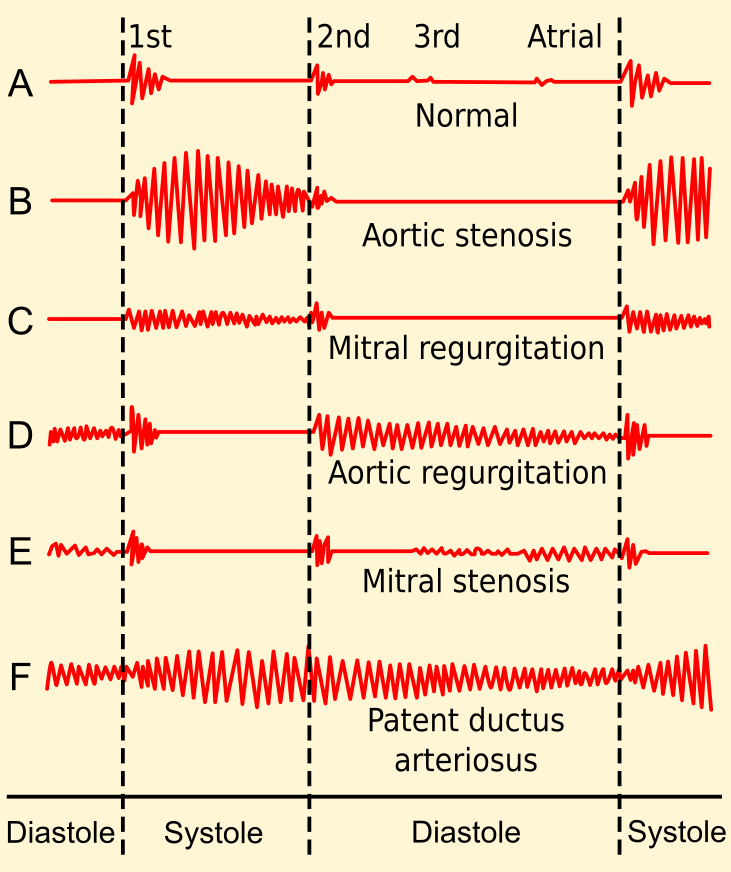
A “blowing or swooshing sound”, “audible when blood volume [is] increased in [the] heart or blood flow is impeded or altered”, from “cardiovascular disease or circulatory problems”. Systolic: just after S1. Diastolic: just after S2. The lab manual: graded 1 to 6; “a diastolic murmur or murmur > grade 3/6 is never innocent”; abnormal aortic murmurs are heard best at Erb’s point.
Original pitch, for headphones
Original pitch, for headphones
The rest of slide 21
| Sound | What the deck says |
|---|---|
| Split sounds | “Normal variation (valves not closing exact same time)” — one sound becomes two. Lab manual: normal split S2 at the pulmonic area, on inspiration; a split S1 at the lower left sternal border possible. |
| Systolic ejection click | “Sudden distention of the great arteries” — early systole, as the semilunar valves open. |
| Opening snap | “Associated with mitral stenosis” — a stiff mitral valve snapping open in early diastole, the exception to “opening is silent”. |
| Pericardial friction rub | Listed without comment — see the next section. |
Original pitch, for headphones
The Think Conceptually handout’s sound list adds muffled; in standard teaching that suggests fluid around the heart (pericardial effusion).
Pericardial or Pleural Rub?
Both are a scratchy, grating sound of two membranes rubbing; the difference is what moves them.
- A pleural rub stops — the visceral and parietal pleura rubbing (Jeopardy’s wording), which move only when the lungs move.
- A pericardial rub keeps going, because the heart keeps beating.
| Pleural rub | Pericardial rub | |
|---|---|---|
| Layers rubbing | Visceral and parietal pleura — around the lungs | Layers of the pericardium — the sac around the heart |
| Keeps time with | Breathing | The heartbeat |
| Client holds breath | Stops | Continues |
| Pain it goes with | The deck’s chest-pain list: “Pulmonary disease / Pleural pain” | The same list: “Pericardial pain” |
Standard teaching: ask for only a brief breath-hold; a pericardial rub is often loudest at the lower left sternal border with the client sitting up and leaning forward.
Bruits and Thrills
Murmur, bruit, thrill: one event found three ways — turbulent flow through a narrowed or damaged opening. Where you find it, and whether you hear or feel it, names it. The Exam IV review: a bruit is “a swishing sound… in the carotid artery or any other artery”.
| Where | Heard or felt | What it means | What you do next | |
|---|---|---|---|---|
| Murmur | Over the heart | Heard — blowing | Turbulence through a valve or chamber | Note timing, site and grade; report a new one |
| Bruit | Over an artery — carotid, abdominal aorta, femoral | Heard, with the bell — “swishing” | Narrowing or disrupted flow | Report — a sign of arterial disease (at the neck, possibly an enlarged thyroid) |
| Thrill | Heart or artery | Felt — a fine vibration | Turbulence strong enough to palpate | Listen there: a murmur over the heart, a bruit over a carotid |
| Pericardial rub | Over the heart | Heard — scratchy | Inflamed pericardium, not flow | Breath-hold — it keeps going |
Original pitch, for headphones
The recorded lecture: “we utilize the bell of our stethoscope to listen for bruits. And that’s a swishing sound.” Jeopardy: a bruit “can indicate a narrowing or disruption of blood flow.” HEENT deck: an enlarged thyroid can disturb carotid flow, so auscultate for a bruit. Cardiac deck notes: carotids one at a time, so the brain’s supply is not cut off.
Standard content: feel a thrill with the palm or base of the fingers; it first appears at murmur grade 4. Lab manual: a bruit can be normal in children and high-output states.
The One Rule: Look One Stop Backwards
The Exam 4 blueprint’s version of the heart lab’s “what happens with abnormalities” adds the clue: “Important to understand blood flow and internal structures to answer these types of questions.” You work the symptoms out from the fourteen stops. Blood is a one-way queue: when a chamber or valve fails, blood waits in whatever comes before it, and whatever comes after it goes short — a backward half (congestion) and a forward half (not enough arriving).
Drag sideways to see the whole figure →
| What fails | Backs up into (look back) | Goes short (look forward) |
|---|---|---|
| Tricuspid valve · 3 | Right atrium (2), the superior and inferior vena cava (1), the body. The prior-year review asks exactly this. | Less reaches the right ventricle and the lungs. |
| Right ventricle · 4 | Right atrium, venae cavae, body — neck veins, legs, liver. | Less reaches the lungs, so in the end less reaches the body. |
| High pressure in the pulmonary artery · 6 | The right ventricle pushes harder, tires and fails; then the cavae back up. | Less blood gets through the lungs. |
| Mitral valve · 10 | Left atrium (9), pulmonary veins (8), lungs (7). | Less reaches the left ventricle and the body. |
| Left ventricle · 11 | Left atrium, pulmonary veins, the lungs. | Decreased cardiac output to the whole body. |
Left- and right-sided failure as named syndromes, and the backward/forward framing, are standard nursing content (Taylor Ch 40; ATI Cardiovascular); this note covers every left-versus-right table in this part.
Left Side Fails — the Lungs Fill
When the left ventricle cannot empty — scarred by an MI, or worn by years of high blood pressure — blood waits in the left atrium, pulmonary veins and lungs, and fluid is pushed into the air spaces: every backward sign is wet lungs.
| Finding | Why it happens — and where the course shows it |
|---|---|
| Dyspnea | Fluid in the air spaces gets in the way of gas exchange. “Difficulty breathing” is on the deck’s bolded history list. |
| Orthopnea | Breathless lying flat, better sitting up — lying down returns fluid from the legs and spreads the lung fluid; sitting up lets it settle. The deck’s question: “How many pillows do you use for sleep?” One pillow to three is the lungs filling. |
| Crackles | Air bubbling through fluid, heard first at the bases. HB (anterior-wall MI on a background of hypertension) has “bibasilar crackles”. The Think Conceptually handout: “Auscultate lung sounds — (pulmonary edema associated with cardiac dysfunction)”. |
| Cough | The airways clearing the fluid; on the deck’s past-history slide beside shortness of breath and orthopnea. |
| Fast, shallow breathing · low SpO2 | Less oxygen crosses at stop 7. HB’s respirations are 28/min, shallow. |
Standard nursing content: the mechanisms in the “why” column; the cough of pulmonary edema can bring up frothy, pink-tinged sputum; some patients wake suddenly at night short of breath (paroxysmal nocturnal dyspnea).
The forward half: the left ventricle is the only pump that feeds the body, so its failure is decreased cardiac output — see Not Enough Forward, below.
Right Side Fails — the Body Fills
When the right ventricle cannot keep up, blood waits in the right atrium, the venae cavae and every vein that empties into them — neck above; liver, gut and legs below. The lungs are downstream, so they can stay clear.
| Finding | Why it happens — and where the course shows it |
|---|---|
| Jugular vein distention (JVD) | The jugulars drain straight into the superior vena cava, so the backup shows there first. Jeopardy: “Bulging of this vein can indicate right sided heart failure”. Handout: “Assess for jugular vein distention (venous congestion from heart failure)”. The deck lists it under inspection. |
| Peripheral, dependent edema | Fluid pushed into the tissues where gravity takes it — feet and ankles up and about, the sacrum in bed — usually both sides. On the deck’s history list and cues. |
| Weight gain | About a kilogram a litre, so the scale shows it before the ankles. Deck cue “Weight Gain”; handout “Assess Daily weights — fluid status”. |
| Enlarged liver, swollen abdomen | The liver drains into the inferior vena cava, so it swells (hepatomegaly) and fluid can collect in the abdomen. HB’s form checks for it — “negative hepatomegaly”. |
Standard nursing content: abdominal congestion can cause loss of appetite and nausea; the abdominal fluid is ascites. The lab manual gives the normal finding as no neck vein distention, jugular venous pressure under 3 cm with the patient at 45° (more in Part 5).
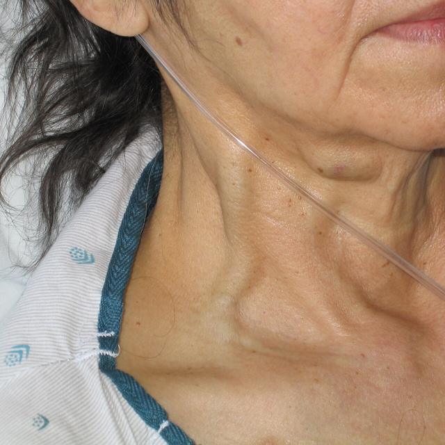
“What assessment finding is associated with tricuspid valve dysfunction (blood backing up from the dysfunctional valve)?” It “would back-up blood flow in the Superior/Inferior Vena Cava”: JVD and peripheral edema.
“Which ventricle’s workload will increase if there is high pressure in the pulmonary artery?” The right — it empties into that artery, so the pressure is its afterload. The handout: “high pressure — heart has to work harder — failure”.
Standard nursing content: chronic lung disease, raising pulmonary artery pressure, is the other classic cause — one reason the deck’s history slide asks about “Lung disease”.
Why Heart Patients Get Up at Night
Nocturia is getting up during the night to urinate — Jeopardy’s “A nurse charts this term when a patient reports having night time urination.” Three steps:
1. By day, the fluid sits in the legs as dependent edema, out of the circulation — the weak heart is not pumping it and the kidneys are not seeing it.
2. At night, lying flat brings it back. The fluid moves from the tissues into the veins, the resting heart pumps it on, and more blood reaches the kidneys.
3. More blood through the kidneys makes more urine overnight — so the patient wakes to urinate, and the ankles are smaller by morning.
The prior-year review: “Nocturia (frequent night urination) and Edema are symptoms of which Heart Condition?” — heart failure. The deck pairs them on its bolded history list and asks “Do you get up during the night to urinate?” right after the pillows question: orthopnea is the lungs’ answer to lying down, nocturia the kidneys’.
The mechanism is standard physiology (Taylor Ch 40). Nocturia has other causes — prostate, diabetes, an evening diuretic, bedtime fluid — so look for it paired with edema.
Not Enough Forward: Decreased Cardiac Output
Cardiac output: Jeopardy’s “how much blood the left ventricle pumps out in a minute”, “a reflection of blood flowing around the body, especially to the brain and other vital organs.”
Standard: resting output is roughly 4–8 litres a minute; the compensating rise in rate costs the heart oxygen.
The short answer is decreased tissue perfusion — the prior-year review’s “How does it affect tissue perfusion?”
| Where | What you find | Source |
|---|---|---|
| Circulation | Blood pressure falls; heart rate rises to compensate; pulses weak (+1). | Jeopardy: BP is “highly reflective of cardiac output”. HB: +1 peripheral pulses. |
| Brain | Dizziness, restlessness, confusion, a change in level of consciousness. | Handout: “Monitor for changes in level of consciousness (blood getting to brain?)”. Dizziness is on the deck’s history list. |
| Kidneys | Less urine; BUN and creatinine rise. | Handout: “BUN, Creat — elevate if impaired blood flow to Kidney as in Heart Failure”; “Monitor Intake and Output”. |
| Skin & extremities | Cool, pale skin, slow capillary refill, weak peripheral pulses. | HB: skin “pale/ashen”, “poor capillary refill”, skin cool. |
| Muscles | Fatigue, tiring easily, unable to keep up with daily activities. | Deck history list: “Fatigue/ tire easily”; lifestyle slide: “Can you perform daily activities?” |
| The heart itself | Chest pain — the heart muscle is short of oxygen too. | Deck: cardiac pain is “cells deprived of O2”. |
Standard: on a nitroglycerin drip, like HB, BP is also what the drug can drop.
| Watching for | Interventions (handout wording) |
|---|---|
| Forward failure — output | Monitor vital signs (temp, pulse rate and rhythm, respirations, BP) · EKG · changes in level of consciousness · labs (Hgb, K, Mg, creat) |
| Backward failure — lungs | Auscultate lung sounds · oxygen as indicated · position for improved ventilation — “increased lung expansion, improve venous return” |
| Backward failure — body | Jugular vein distention · peripheral edema · intake and output · daily weights |
| The heart itself | Heart sounds (extra sounds, murmur, muffled, rub) · assess chest pain, treat promptly · bedrest if acute MI or CHF exacerbation — “decrease workload of heart” — then progressive activity |
Left-Sided Versus Right-Sided, Side by Side
Sort a stem’s findings into the columns; whichever fills is the failing side. Both? The left probably went first.
| Left side fails | Right side fails | |
|---|---|---|
| The failing pump | Left ventricle (11), or the mitral valve (10) before it | Right ventricle (4), or the tricuspid valve (3) before it |
| Backs up into | Pulmonary veins → lungs | Venae cavae → body |
| Lung sounds | Crackles, at the bases first | Usually clear — unless the left side is failing too |
| Breathing | Dyspnea, orthopnea, cough, fast shallow respirations, low SpO2 | Not the main complaint |
| Neck veins | Flat, at first | JVD |
| Edema & weight | Not the first sign | Dependent edema, both legs or the sacrum · weight gain |
| Abdomen | — | Enlarged liver, swollen abdomen |
| Forward shortfall | Decreased cardiac output to the body: falling BP, fatigue, dizziness, LOC changes, cool pale skin, less urine | Less blood to the lungs, and so in the end to the left heart and the body |
| The deck’s question that finds it | “How many pillows do you use for sleep?” | “Edema or swelling” |
| Either side | “Do you get up during the night to urinate?” — nocturia points to heart failure, not to one side | |
| Stem words that point here | lungs, crackles, short of breath, pillows, sits up to breathe, cough, mitral | neck veins, JVD, ankles, swelling, weight up, liver, tricuspid, cavae, pulmonary artery pressure |
| Usual cause | MI, long-standing high blood pressure (HB has both) | Left-sided failure; lung disease; high pulmonary artery pressure |
- Left, backward: bibasilar crackles; respirations 28, shallow.
- Left, forward: skin pale/ashen, cool, poor capillary refill, +1 peripheral pulses; heart rate 115.
- Right: “Jugular Vein Distention Note[d]” — but negative hepatomegaly, no edema recorded.
- Heart sounds: a positive S3.
HB’s case gives both “poor capillary refill” and “< 3 seconds”, so build no answer on his refill time. That an anterior-wall MI involves the left ventricle (left anterior descending artery) is standard anatomy, not stated in the case.
Traps
The Cardiac History
The cardiac deck spends four slides on subjective data (slides 10–13) before it reaches for the stethoscope, and that proportion is the lesson: most of what the exam will later confirm, the history has already found. Crackles, JVD and an S3 are what you hear and see; orthopnea, nocturia and “I can’t keep up any more” are what the patient tells you first. Parts 3 and 4 explain the findings. This part is the conversation that leads to them.
History of present concern — the deck’s bolded list
Slide 10 sets every line of this list in bold. Learn it as a list of cues, each pointing at a part of the heart’s job:
| Symptom (deck wording) | What it points to | Explained in |
|---|---|---|
| Pain… angina (chest, arm, jaw) | Heart muscle short of oxygen — “cardiac pain… cells deprived of O2” (slide 14). | The next section |
| Irregular (skip) beats / palpitations | A rhythm problem — the conduction system, not the pump. The patient’s awareness of the beat, so it is subjective even though it sounds like a measurement. | Part 3 (rate and rhythm, pulse deficit) |
| Fatigue / tire easily | Forward failure — muscles not getting enough blood. | Part 4 |
| Difficulty breathing | Backward failure into the lungs — the left side. | Part 4 |
| Orthopnea | Breathless lying flat, better sitting up — left-sided. Measured by the pillow question below. | Part 4 |
| Dizziness | The brain under-perfused — low output or a rhythm problem. | Part 4 |
| Nocturia | Fluid from swollen legs returning to the circulation at night — heart failure. | Part 4 |
| Edema | Venous backup — right-sided failure, or the leg’s own veins. | Parts 4, 6 and 7 |
| Heartburn / indigestion | Can be cardiac pain wearing a GI label — HB’s own word for his MI. | This part |
| Diaphoresis / sweating | The body’s alarm response. Sweating alongside chest pain is a cardiac cue until proven otherwise. | This part |
Standard nursing content: diaphoresis in an MI is a sympathetic (“fight or flight”) response to pain and falling output; the deck lists it without explaining it.
Demographics, past history and family history — the speaker notes
Slide 10’s notes and slide 11 give the rest of the history. Demographics first: age, gender, baseline pulse and BP, recent blood work — the baselines you will compare everything else against.
| Area | What the deck has you ask |
|---|---|
| Heart problems | Ever diagnosed with chest pain, heart attack, heart failure or an irregular rhythm? A murmur? Rheumatic fever? |
| Blood pressure | Has anyone told you that you have high blood pressure? What is your normal range? Do you take anything for it? Do you monitor your own BP and HR? |
| Cholesterol | Has anyone told you that you have high cholesterol? What is the usual level? Do you take anything for it? Have you had a lipid profile? |
| Diabetes | History of diabetes mellitus? Usual glucose level? Do you take anything for it? |
| Other conditions | Pre-existing diabetes, lung disease, HTN. |
| Past tests | Had an ECG? When? Results? Blood tests? |
| Family history | Any family history of cardiovascular problems? Then five follow-ups: who had it, what was it, when did they have it, how was it treated, what was the outcome? |
Why the family-history follow-up asks when: heart disease in a parent or sibling at a young age is a stronger risk signal than the same disease at 85 (standard nursing content). HB’s father died of an MI at 57.
Medications — including the ones they buy themselves
Slide 11: “Medications for heart disease? Why? Assess compliance” and “OTC meds and nutritional supplements?” Slide 13’s notes add the reason: “OTC meds/supplements (some can interact with prescribed meds)”, and ask for the medication schedule as well as the names. Three separate questions hide in that line: what do you take, why do you take it (the answer often names a diagnosis the patient forgot to mention), and do you actually take it as prescribed.
Standard examples of OTC products that matter to a cardiac patient: decongestants and NSAIDs can raise blood pressure, and some herbal supplements interact with anticoagulants.
Lifestyle and health practices — slide 12
| The question | What it is really asking |
|---|---|
| Do you smoke? | The biggest modifiable risk. The notes add how long and how much. |
| What type of stress do you have? How do you cope? | Stress is on the handout’s risk list and the review sheet’s “reducing heart failure risk” list. |
| Describe what you eat in a 24 hour period? | Diet — asked as a recall of real meals, not “do you eat healthily?”, which everyone answers yes. |
| How much alcohol do you drink? | Notes: how long, how much, what types. |
| Do you exercise? What type and how often? | Activity level — slide 9’s goal is more adults doing vigorous activity. |
| If heart disease… has it impacted sexual activity? | Function and quality of life; asked plainly, as part of the routine. |
| Can you perform daily activities? | Fatigue and forward failure, and the functional baseline. |
| How many pillows do you use for sleep? | Orthopnea — a left-sided failure screen (Part 4). |
| Do you get up during the night to urinate? | Nocturia — a heart-failure screen (Part 4). |
| What fears do you have? | Anxiety, which adds to the heart’s workload — HB asks, “Am I going to die?” |
Slide 13’s notes add three more: usual weight, typical diet, and recreational drug use.
Chest Pain: Ask, Assume, Act
The first question: have the client describe the pain. The review sheet is unusually emphatic: “it is most important to have them describe the pain and know what it feels like for the patient… Other information is important. However, this is the most important and first additional information you want to know.” Not “have you had this before?”, not “what did you eat?”, not the pain scale on its own — “tell me what it feels like.”
Where it is, and what it is called: the deck’s history list says “Pain… angina (chest, arm, jaw)”, and the review sheet: “chest pain (and any radiating pain) is known as angina.” Jeopardy defines it twice — “chest pressure that can be stable or unstable” and “pain [that] occurs with a reduction of oxygen to the heart”. The deck’s own one-line definition: “cardiac pain… cells deprived of O2.”
Start hides the labels and names a place — click where it is on the patient. Remember the figure faces you, so the patient’s left arm is on your right.
Drag sideways to see the whole figure →
Stable versus unstable angina, standard nursing content (Taylor Ch 40 defines stable angina as a temporary imbalance between the oxygen the heart needs and the oxygen delivered): stable angina is predictable — brought on by exertion, eased by rest or nitroglycerin. Unstable angina is new, worsening, or comes at rest, and is treated as an emergency. Pain that lasts longer and is not relieved points toward an MI.
What to ask about chest pain — a checklist
Once the patient has described the pain, the rest of the questions fill in the picture. OPQRST is a common way to remember them. It is a study aid, not course wording — the course’s own rule is only that the description comes first, so start at Q and use the letters to make sure nothing is missed. HB’s answers, from the cardiac case, are in the third column.
| Letter | Ask | HB said | Why it matters |
|---|---|---|---|
| Quality — first | “Describe it to me. What does it feel like?” | “Feels like someone sitting on my chest — pressure!” | Pressure, squeezing, heaviness are the cardiac words. The review sheet’s “first” question. |
| Region & radiation | “Show me where. Does it go anywhere else?” | “Right here in the middle of my chest.” Radiation not recorded. | Central chest is the classic site. Ask about the arm and jaw — the case does not say. |
| Severity | “On a scale of 0 to 10?” | 10/10 | A number to re-measure after treatment — it becomes your evaluation. |
| Onset | “When did it start? What were you doing?” | About half an hour after a large dinner; the pain woke him from a nap. | Pain that comes at rest or wakes someone is not the predictable, exertional pattern of stable angina. |
| Provokes / palliates | “What makes it worse? Better? What have you taken for it?” | Took antacids — “nothing seemed to make it better.” | Failed relief is a cue. So is what he took: he had already labelled it. |
| Time | “How long has it lasted? Constant? Ever had it before?” | About two hours. “I never had indigestion this bad before.” | Long-lasting, and different from his usual — a change in pattern is a cue in itself. |
| Associated symptoms | “Any sweating, shortness of breath, nausea, dizziness?” | Diaphoretic (observed). Denies shortness of breath at rest. | The deck’s history list again. Note he denies breathlessness while breathing 28, shallow — see the HB case below. |
OPQRST (onset, provocation/palliation, quality, region/radiation, severity, time) is a standard pain-assessment mnemonic, not from the course documents; Taylor’s respiratory history asks the same things (when did it start, where, 0–10, worse with breathing or activity).
The only red line in the cardiac deck, at the foot of its chest-pain differential. The list names seven causes, and the red line tells you which one to assume while you find out. The right-hand column below is how the others are usually told apart — and the red line means none of those clues lets you skip the cardiac work-up.
| Cause | In the course documents | What usually points to it |
|---|---|---|
| Myocardial infarction | First on the list | Pressure that lasts and is not relieved by rest or nitroglycerin; sweating, nausea, fear. |
| Angina (stable or unstable) | “Cardiac pain… cells deprived of O2” | Stable: with exertion, eased by rest. Unstable: at rest, new or worsening. |
| Gastrointestinal disease | Handout: indigestion “can be associated with MI” | Burning, linked to meals, eased by antacids. HB had all three cues and an MI. |
| Pulmonary / pleural pain | “Pulmonary disease / Pleural pain” | Sharp, worse on breathing in; a pleural rub that stops when the breath is held (Part 3). |
| Musculoskeletal | “Musculoskeletal issues” | Tender to touch, worse with movement of the chest or arms. |
| Aortic dissection | “Aortic Dissection” | Sudden, severe, tearing pain, often through to the back. |
| Pericardial pain | “Pericardial pain” | Sharp, eased by sitting up and leaning forward; a pericardial rub that carries on when the breath is held (Part 3). |
The first minutes, in order
Putting the review sheet, slide 23, Jeopardy and the handout together. Every step is correct; the question an exam asks is which comes first.
| # | Step | Source and reason |
|---|---|---|
| 1 | Assume it is cardiac | Slide 14 red text. It decides how fast everything else happens. |
| 2 | Have the patient describe the pain | Review sheet: “the most important and first additional information.” |
| 3 | Vital signs — BP above all | Jeopardy: BP is the priority with chest pain. (Standard: nitroglycerin lowers BP, so a BP is needed before it is given.) |
| 4 | Oxygen and nitroglycerin as ordered, while gathering the rest | Slide 23: “gather data while treating.” Not after. |
| 5 | Notify the provider | Review sheet: “Is it important to notify the MD about this pain?” — yes. Handout: “treat promptly if present.” |
| 6 | Reassess the pain score and BP | The 10/10 becomes the measure of whether treatment is working. |
| 7 | Document | Last, as always. |
The order is a synthesis, not a list any course document prints. In practice steps 2–5 overlap in the first minute, and standing orders or an emergency protocol may start oxygen before you have finished asking. On a paper question, “describe the pain” wins the what do you ask first item, and BP wins the which vital sign item.
Standard nursing content, not spelled out in the deck: women, older adults and people with diabetes more often have “atypical” symptoms — unusual fatigue, shortness of breath, nausea, indigestion, back, neck or jaw pain — and older adults may present with confusion or breathlessness alone.
Risk Reduction and Teaching That Works
Slide 13 is titled “Risk assessment and health-related patient teaching (Address Modifiable Risk Factors)” and its notes say “Stress patient education for modifiable risk factors identified during subjective assessment.” So the history above is where the teaching plan comes from: every risk factor the patient tells you about is a teaching topic. Sort them first:
| Modifiable — teach to these | Non-modifiable — screen and watch |
|---|---|
| Smoking — slide 13’s first line | Genetics |
| Blood pressure / hypertension — control it | Family history of heart disease |
| Cholesterol — hyperlipidemia (“fat in blood”) | Age |
| Overweight and obesity | Gender |
| Elevated glucose — diabetes control | Ethnicity |
| Physical inactivity (“sedentary”) and diet | |
| Stress; caffeine (the handout: “caffeine intake — dysrhythmia”); alcohol; recreational drugs |
The prior-year review asks it another way — “What are some recommendations for reducing Heart Failure Risk? Manage Stress? Blood Pressure? Smoking?” All three are yes, and all three are on the modifiable side.
The population goals — slide 9
Slide 9 states the public-health goal as “Reduce CHD deaths”, and lists how: smoking cessation; educate adults on early warning signs and symptoms — call 911; increase timely therapy (an AED in the community); increase HTN control; reduce cholesterol; more medical office visits for people with co-morbid conditions such as diabetes and high cholesterol; and more adults doing vigorous physical activity.
Standard teaching that goes with “call 911”: do not drive yourself to hospital with chest pain, and do not wait to see whether it passes.
Who carries more risk — slide 24
| Group | What the deck says |
|---|---|
| Everyone | Heart disease is the leading cause of death in high-income countries, and across all races and ethnicities in the US. |
| Older adults | Age-related changes: stiffening of the vasculature, hypertrophic ventricles, atherosclerotic disease, valve disease. |
| Women | Present differently than men; underrepresented in research. |
| African American men | Greater risk of cardiovascular disease and stroke. |
Teaching that motivates
This reading of the review-sheet question is an inference from the Jeopardy answer, not a printed course answer. It matches standard nursing teaching principles (readiness to learn; motivational interviewing): fear-based messages alone tend to produce short-lived change.
The Physical Exam: Prepare, Inspect, Palpate
Slide 15 is two questions — “What equipment is needed? How will the nurse prepare the patient?” — with the answers set to “fly in” one at a time. A slide built as a quiz is a good guess at an exam question.
| What the deck lists | |
|---|---|
| Equipment | Stethoscope; watch with a second hand. |
| Preparation | Explain the procedure · pull the curtain / close the door · patient in a gown · expose only the part of the chest being assessed, “especially for women” · provide a blanket or sheet for warmth and privacy. |
Slide 16 then names the techniques, in the usual order — inspect, palpate, percuss, auscultate:
| Technique | What you look or feel for | Expected, and the deck’s notes |
|---|---|---|
| Inspection | Lesions; masses; deformity; jugular vein distention; PMI. Notes add areas of tenderness. | PMI may be visible at the apex — easier in children, men, and thin-chested adults (slide 17: “can see on child 1 cm elevation”). JVD is the right-sided backup sign of Part 4. |
| Palpation | PMI; carotid pulse; masses. | Use the finger pads in the left 5th ICS, midclavicular line. The PMI “may or may not be palpable in adults.” No pulsations are felt anywhere else. The lab-manual table adds: note thrills, lifts or heaves — “If you palpate a thrill, listen for a murmur” (Part 3, Bruits and Thrills). |
| Percussion | Cardiac borders — “(Advanced)”. | “Not commonly performed.” |
| Auscultation | Rate and rhythm, then S1, S2 and extra sounds at the five sites. | All of Part 3 — APETM, base and apex, gallops, murmurs, rubs. The lab-manual table: “Auscultate in sitting, supine, and left lateral recumbent positions”, with the bell (light pressure) and diaphragm (heavy pressure) at all sites; the deck: S3 is best heard in the left lateral position. |
Standard nursing content: a PMI that is displaced to the left of the midclavicular line, or larger than about a coin, suggests an enlarged left ventricle — the left ventricular hypertrophy HB’s old ECG showed.
What it looks like written down. HB’s focused assessment is a model of the format: “Neck vessels: positive carotid pulsations, +2 symmetrical. Jugular vein distention noted.” and “Precordium: pulsation noted at point of maximum impulse. Heart sounds: tachycardia, irregular rhythm. S1 > S2 at apex, S2 > S1 at base, positive S3 noted.” Every phrase in it is something this part or Part 3 taught you to look for.
From the Lab-Manual Table: Neck Vessels and Percussion
The lab manual’s cardiovascular assessment table adds three things the deck only names: how to tell a carotid pulsation from a jugular one, a pressure test on the neck veins called abdominojugular (hepatojugular) reflux, and what percussion of the heart finds.
| Carotid (artery) | Jugular (vein) | |
|---|---|---|
| The wave | One positive wave — a single outward beat | Undulating wave — a soft, rippling rise and fall |
| Light pressure | (Standard: still felt — it is a palpable pulse.) | Easily obliterated — it disappears |
| Position and breathing | (Standard: unchanged.) | Affected by position and respirations |
| Normal finding | Visible carotid pulsation; palpated one side at a time, +2 | No neck vein distension. Jugular venous pressure, measured with the patient at 45° from the sternal angle (angle of Louis): under 3 cm |
Abdominojugular (hepatojugular) reflux. The table’s palpation row: “Palpate jugular veins and check direction of fill. Check for abdominojugular (hepatojugular) reflux.” The expected finding is “Jugulars easily obliterated and fill appropriately. Negative abdominojugular reflux.” Negative is normal.
Standard nursing content (the table names the test but not the method): with the patient at 30–45°, press firmly over the right upper abdomen, over the liver, for about 10 seconds and watch the neck veins. A jugular pressure that rises and stays up while the pressure is held is a positive reflux: the right side of the heart cannot take the extra blood pushed back to it. That points to right-sided heart failure — the same backup that causes JVD in Part 4.
Percussion of the heart. Slide 16 lists “Percussion (Advanced) — Cardiac Borders — not commonly performed”, so you will not be expected to do it. The lab-manual table says how it is done and what is normal: indirect (mediate) percussion, from the anterior axillary line toward the sternum at the fifth ICS; expected finding, dullness at the third, fourth and fifth ICS to the left of the sternum. The heart is solid, so it sounds dull where the air-filled lung around it sounds resonant — the change from one to the other marks the heart’s border.
Standard nursing content: dullness that reaches further left than expected suggests an enlarged heart. In practice a chest X-ray or echocardiogram answers that question, which is why the deck calls percussion advanced and rarely done.
Labs and Diagnostic Tests
Three of the blueprint’s cardiovascular lines are about tests: the lipid profile, both kinds of echocardiogram, and preparing for a stress test. They get a blue box each. Everything else on slide 22 and the handout’s list is in the tables, so you can recognise it in a stem.
The slide 22 notes: “Elevated levels of blood lipids are a risk factor for cardiovascular disease. A lipid profile includes total cholesterol, high-density lipoprotein (HDL), LDL, and triglyceride levels.” The targets, with the one capital letter in the notes kept:
| Test | Target | Remember it as |
|---|---|---|
| Total cholesterol | < 200 | “Desirable.” A normal total can hide a high LDL. |
| LDL | < 100 | “Optimal.” The “bad” cholesterol (Jeopardy) — and the review sheet’s answer to “which cholesterol is the most dangerous for heart disease?” |
| HDL | GREATER than 40 | The good one. The deck capitalises GREATER because it is the only target you want high. |
| Triglycerides | < 150 | The other fat in the blood. |
Drag sideways to see the whole figure →
A study aid, not course wording: LDL = Lousy (keep it Low); HDL = Healthy (keep it High). Slide 22 also asks “Patient education?” for the lipid profile. Standard teaching: the patient may be asked to fast beforehand (traditionally 9–12 hours; follow the lab’s instruction); a diet low in saturated fat, weight loss and medication lower LDL; regular exercise raises HDL and smoking lowers it.
Blood tests
| Test | What it tells you |
|---|---|
| Cardiac markers — troponin-I, CK-MB, myoglobin | “The myocardium releases cardiac enzymes and proteins in response to cell damage… measured in blood samples to diagnose or rule out MI.” CK-MB and troponin-I are “specific to the myocardium; their values are elevated with MI.” Slide 22 lists total CK and CK-MB and troponin-I; the handout adds myoglobin. HB had a “serum cardiac enzyme test” drawn in the ED. |
| BNP — B-type natriuretic peptide | “Marker for heart failure.” |
| Electrolytes (Na, K, Mg, Ca) | “Affect electrical and ventricular function.” Abnormal potassium or magnesium is a rhythm risk. |
| BUN, creatinine | “Elevate if impaired blood flow to kidney as in heart failure” — Part 4. |
| CBC | “Volume / Hgb / WBC infection.” |
| Blood coagulation factors | Clotting — checked when anticoagulants are given. |
| Serum lipids | “Fats” — the lipid profile above. |
| Homocysteine | “Elevation indicates risk for CV disease.” |
| C-reactive protein (CRP) | “Detects inflammatory process.” |
Approximate timing, standard nursing content: myoglobin rises first (within a few hours) but is not specific to the heart; CK-MB rises within hours and returns to normal in two to three days; troponin rises within hours and stays raised for one to two weeks, so it can confirm an MI in a patient who arrives days later. Serial draws are usual. BNP is released by stretched ventricles — the higher it is, the worse the failure.
Diagnostic tests
| Test | What it shows | What you teach / prepare |
|---|---|---|
| Electrocardiogram (ECG, EKG) | “Tracing of electrical conduction.” Helps diagnose ischemia, chamber hypertrophy, pericarditis, electrolyte imbalances, arrhythmias, heart block. Jeopardy: an arrhythmia is “an electrical malfunction of the heart”, and asystole — “sometimes called flatline” — “results in no cardiac output.” | Electrodes on the limbs and front of the chest with adhesive pads (deck). *Painless; lie still and breathe normally; no electricity goes into the body. |
| Holter monitor | “Noninvasive, monitor ECG over prolonged period.” | *Worn at home, usually 24–48 hours. Keep a diary of activities and symptoms with times; keep the device dry. |
| Chest X-ray | “Size, contour, and position of the heart; alterations in the pulmonary circulation; acute or chronic lung disease.” | *Remove metal from the chest; ask about possible pregnancy. |
| Echocardiogram — transthoracic or transesophageal (TEE) | “High frequency sound waves and the Doppler effect” — size, shape and motion of atria, ventricles and valves, and the direction and velocity of blood flow. | The blue box below. |
| Stress test — exercise or chemical | “Compares cardiac function and perfusion at rest versus during stress.” | The blue box below. |
| Cardiac catheterization | “To diagnose the presence of coronary artery blockages.” | *Invasive: consent; NPO beforehand; ask about contrast-dye allergy and kidney function. Afterwards: check the insertion site for bleeding and the pulses below it, keep the limb straight as ordered. |
| Electrophysiology (EP) studies | On slide 22 and the handout; the deck does not explain them. | *Catheters map the heart’s electrical conduction and may provoke a dysrhythmia on purpose; prepared like a catheterization. |
| Nuclear studies (thallium scan) | On the handout list. | *A small dose of radioactive tracer shows which areas of muscle are perfused; often paired with a stress test. |
| Cardiac MRI | On the handout list. | *Screen for metal and implanted devices (pacemaker); lie still in an enclosed scanner. |
| Hemodynamic monitoring | On slide 22; not explained in the deck. | *Pressures measured through lines placed in an artery or the heart; an intensive-care tool. |
The review sheet asks it as two questions: “If a patient is getting a transthoracic echocardiogram (an ultrasound of the heart) what does this procedure entail? How is this different from a transesophageal echocardiogram?” Both use the same thing — ultrasound. The difference is where the transducer goes.
| Transthoracic (TTE) | Transesophageal (TEE) | |
|---|---|---|
| Where the transducer goes | On the chest wall. “A gel (often cold) is placed on the patient’s chest wall and the transducer is moved around the anterior chest wall.” | Down the oesophagus on a flexible probe, so it sits directly behind the heart. |
| Invasive? | No. Painless. | Yes — a scope is swallowed. |
| Before | Explain it; warn that the gel is cold; gown, privacy, lie on the back or left side. | NPO for several hours; consent; IV access; dentures out; the throat is numbed and the patient is sedated. |
| After | Wipe off the gel; nothing else. | Nothing to eat or drink until the gag reflex returns (aspiration risk); monitor vital signs and sedation; a sore throat is expected; someone else drives home. |
| Why choose it | The first-line look at size, motion, valves and flow. | A clearer picture when ribs, lungs or body size block the chest view, and of structures at the back of the heart such as the left atrium (clots) and valves. |
Drag sideways to see the whole figure →
A stress test “compares cardiac function and perfusion at rest versus during stress” (slide 22 notes), and the handout says it is exercise or chemical. The whole point is to make the heart work harder — to push the heart rate up — and see whether the muscle still gets enough blood. So a drug that holds the heart rate down defeats the test. That is the blueprint’s answer: the meds to hold are the meds that slow the heart rate, and the classic one is the beta blocker. The review sheet asks it as “Will medications that effect HR be held?” — yes.
| Teach | Why |
|---|---|
| Hold heart-rate-slowing medications — as the provider directs | So the heart rate can rise. Jeopardy’s wording is “consult with physician about how to adjust prescription medications” — the nurse teaches the provider’s instruction; the nurse does not decide it. |
| No smoking / tobacco or caffeine products before the test | Jeopardy. Both change the heart rate and blood pressure. |
| Wear comfortable clothing | Jeopardy — and walking shoes for a treadmill test. |
| Expect HR, BP and ECG to be watched throughout | The review sheet asks “what VS are we concerned with?” — the heart rate (it must reach its target) and the blood pressure (it should rise with exercise; a fall is a danger sign). |
Standard nursing content: other rate-slowing drugs that may be held are calcium channel blockers such as diltiazem and verapamil, and digoxin; beta-blocker names usually end in -olol (metoprolol, atenolol). A chemical stress test gives a drug that mimics exercise for a patient who cannot walk a treadmill; caffeine is avoided for 12–24 hours because it blocks some of those drugs. The test is stopped for chest pain, a falling BP, a serious dysrhythmia or severe breathlessness. The review sheet prints no answer to its VS question; “HR and BP” is this guide’s reading.
From Cues to Care
The deck closes on the clinical judgement model, with cardiac examples at each step. The case sheet for HB asks you to walk the same steps, so learn them as a sequence:
| Step | Slide 25 |
|---|---|
| 1 · Identify cues | Irregular rhythm; tachycardia or bradycardia; weight gain; JVD; edema; dyspnea; crackles in lungs; murmurs; etc. |
| 2 · Analyse cues and form hypotheses | The common cardiac problems: impaired (decreased) cardiac output; impaired tissue perfusion, cardiac; excess fluid volume. |
| 3 · Prioritise hypotheses | “Includes additional problems discovered during assessment.” |
| 4 · Generate solutions | “What am I going to do about the problems? What do I want to achieve? Goals. Plan to treat and prevent.” |
| 5 · Take action | Interventions. |
| 6 · Evaluate | “Evaluate and modify as needed.” |
How the three problems map onto forward and backward failure is in Part 4, along with the handout’s monitoring interventions (vital signs, ECG, LOC, lung sounds, oxygen, positioning, JVD, edema, I&O, daily weights, bedrest). This section adds the handout’s framework and the interventions Part 4 does not cover.
What a normal heart depends on
The handout opens with a list that works as a diagnostic checklist in reverse: each thing a normal cardiovascular system depends on has a matching way to fail.
| Depends on (handout wording) | Fails through |
|---|---|
| Intact electrical conduction — “maintain rate and rhythm” | Changes in conduction (aging, scar tissue, intrinsic irritability); caffeine → dysrhythmia |
| Circulating blood volume | Too little (bleeding, dehydration) or too much (fluid overload) |
| Patent coronary arteries — “source of oxygen to muscle” | Coronary artery disease / atherosclerosis → MI; hyperlipidemia |
| Intact internal structures | Atrial or ventricular septal defects; congenital problems |
| Normal, healthy heart valves | Valvular disease — prolapse, stenosis, stretched, infection/vegetation |
| Healthy peripheral vasculature — “high pressure — heart has to work harder — failure” | Hypertension; peripheral arterial and peripheral vascular disease (Parts 6 and 7) |
| Proper function of the myocardium — “strength, without restriction” | Scar tissue after MI; hypertrophy of the ventricle; myocarditis, endocarditis; idiopathic |
The first row, in the deck’s words. Cardiac deck slide 7, “Cardiac Conduction System”: the notes say “Electrical impulses fire from the SA node to generate a heartbeat. Essentially the conduction system tells the mechanical system when to ‘beat’.” So the SA node starts each beat, the electrical system sets the rate and rhythm, and the muscle and valves do the pumping. An ECG is the tracing of that electrical conduction. The notes add that the detail comes in the second year, with arrhythmias; for now, know that the beat starts at the SA node.
Standard anatomy: the SA (sinoatrial) node sits in the wall of the right atrium and is the heart’s natural pacemaker. The impulse spreads across both atria, then through the AV node and down the ventricles, so the atria contract just before the ventricles.
Interventions not covered in Part 4
The handout’s collaborative broad goal is “to prevent dysfunction — preserve or maximize the heart function,” with interventions individualised to the patient. Beyond the monitoring list in Part 4, it adds:
- Assess chest pain (cardiac pain) — treat promptly if present.
- Educate the patient regarding the importance of reporting chest pain — a patient who says nothing because “it’s only indigestion” is HB.
- Assess GI — indigestion: “can be associated with MI — plan will be to rule out cardiac issue followed by GI work up.”
- Educate — lifestyle modification: smoking cessation, diet, stress management, medications, activity.
- Administer medications as ordered — below.
Medications — learn them by class
The handout’s advice is the method: “Best to learn medications by classification in order to understand what the RN should be evaluating before and after administering.” Pharmacology comes later in the programme; for now, know the classes on the list and the one thing to check for each.
| Class (handout) | Examples | What the nurse checks |
|---|---|---|
| Diuretics — loop, potassium-sparing | Furosemide; spironolactone | BP, I&O, daily weight, potassium (loop lowers it, potassium-sparing raises it) |
| Antiplatelet | Aspirin (HB, in the ED) | Bleeding |
| Anticoagulants | Heparin, Coumadin (handout) | Bleeding; clotting labs |
| Antiarrhythmic · beta blockers · calcium channel blockers | Metoprolol; diltiazem | Apical pulse and BP before giving — these slow the heart (and are the stress-test holds) |
| ACE inhibitors | Enalapril (HB’s home medication) | BP; potassium; a dry cough |
| Anti-anginal | Nitroglycerin (HB’s drip) | BP — it can drop it; headache; the pain score |
| Antihypertensive | Several of the classes above | BP; dizziness on standing |
| Electrolyte supplements | Potassium, magnesium | The level they replace (handout: “monitor labs — Hgb, K, Mg, Creat”) |
| Thrombolytic | — | Handout: “risks associated determine use” — the risk is bleeding |
The handout’s last page lists exemplars (coronary artery disease, MI, heart failure, cardiogenic shock, valve disorders, endocarditis, myocarditis, pericarditis) and procedures (percutaneous coronary interventions such as angioplasty and stents, valve repair, bypass grafting, pacemakers, implanted defibrillators, ablation) and says they “will be discussed in conjunction with appropriate exemplars in the NUR courses.” They are not on the Exam 4 blueprint.
Worked Case: HB
HB, 72, presents to the ED with chest discomfort that began after dinner and woke him from a nap. Treated for hypertension for four years. Given aspirin and started on a nitroglycerin drip; ECG and cardiac enzymes drawn; admitted to the cardiac care unit with an anterior wall MI. He asks, “Am I going to die?” The case sheet’s reverse side walks the clinical judgement model. Part 4 sorted his findings into left- and right-sided failure; here is the whole case, every cue tied to where this guide explains it.
Step 1 — identify the cues
| Cue | S/O | What it suggests | Explained in |
|---|---|---|---|
| “Terrible chest pain”, “pressure”, middle of the chest, 10/10 | S | Angina / MI — muscle short of oxygen | This part, chest pain |
| “Thought it was indigestion”; antacids did not help; never this bad | S | Cardiac pain in a GI disguise; failed relief; change in pattern | This part, chest pain |
| “Am I going to die?”; affect anxious | S / O | Fear and anxiety — slide 12’s “What fears do you have?” | This part, history |
| Denies shortness of breath at rest | S | His report — but see the next line | — |
| Respirations 28, shallow; bibasilar crackles | O | Fluid backing into the lungs — left-sided failure | Part 4 |
| HR 115, “occasional extra beat”, irregular rhythm | O | Compensating tachycardia; a rhythm problem — count the apical pulse a full minute, check for a pulse deficit | Parts 3 and 4 |
| BP 170/105 | O | High (long-standing HTN, pain, anxiety). The vital sign to keep watching — and the nitroglycerin can drop it | Part 4 |
| Positive S3 | O | Heart failure / fluid overload — “rarely normal in older adults” | Part 3 |
| JVD noted | O | Venous backup reaching the right side | Part 4 |
| Skin pale/ashen, diaphoretic, cool; mucous membranes pale grey; poor capillary refill; +1 pulses | O | Decreased tissue perfusion — forward failure | Part 4 |
| Lower legs cool, pale, shiny and hairless | O | Chronic arterial insufficiency — not the MI | Parts 4 and 7 |
| Sitting upright, clutching his chest | O | Pain; sitting up eases breathing (orthopnea) | This part; Part 4 |
| Temp 100 °F | O | A low-grade rise (standard: common in the first days after an MI as damaged muscle causes inflammation) | — |
| Alert, oriented × 4 | O | Brain still perfused — the baseline for the LOC checks to come | Part 4 |
| Old ECG: left ventricular hypertrophy | O (record) | A left ventricle thickened by years of high pressure | Part 4 |
| 6 ft, 275 lb; HTN for years; large meals | O / S | Modifiable risk: weight (a BMI of about 37), blood pressure, diet | This part, risk |
| Age 72, male; father MI (died at 57), mother stroke, HTN in the family | S | Non-modifiable risk: age, gender, family history | This part, risk |
Two negatives on HB’s sheet. The case also records “negative clubbing” (with the skin) and “negative hepatomegaly” (with the abdomen). A negative is a finding too: it tells you what has not happened yet. Both are signs of a problem that has gone on for a long time, and HB’s problem is sudden.
Standard nursing content: clubbing is a bulbous swelling of the fingertips, with the angle between the nail and the nail fold flattened to 180° or more. It develops over months from chronic hypoxia — long-standing heart or lung disease — so it is not expected in an acute MI. An enlarged liver (hepatomegaly) comes from long-standing right-sided backup, the same congestion that gives a positive hepatojugular reflux.
Steps 2 to 6 — the case sheet’s questions
The case sheet is blank; these are one defensible set of answers built from the course documents, not an answer key.
| The sheet asks | Answer |
|---|---|
| Areas of major concern that warrant continued assessment | Chest pain and the heart itself (pain, rhythm, S3) · breathing (crackles, RR 28) · perfusion (BP, pulses, skin, LOC, urine output) · fluid (JVD, lungs, weight, edema) · anxiety. |
| Explain jugular vein distention | Blood backing up from the right side of the heart into the venae cavae and the neck veins — venous congestion from heart failure (handout). Part 4. |
| What could the S3 indicate? | Heart failure and fluid overload — rapid early filling of a ventricle that is already too full. Rarely normal at 72. Part 3. |
| Hypotheses — actual | Decreased cardiac output and impaired tissue perfusion, cardiac (the MI, pain, HR 115, irregular rhythm, ashen cool skin, +1 pulses) · excess fluid volume (crackles, S3, JVD) · acute pain (10/10) · anxiety / fear (“Am I going to die?”). |
| Hypotheses — risk for | Further fall in cardiac output (the irregular rhythm, the damaged muscle); impaired gas exchange as the lungs fill; bleeding (aspirin and likely anticoagulants); later, a knowledge need about his modifiable risk factors. |
| Prioritise | 1. Decreased cardiac output / impaired cardiac tissue perfusion — the heart muscle is dying now, and the 10/10 pain is that ischemia. 2. Excess fluid volume with its effect on breathing. Pain and anxiety are treated alongside, because both raise the heart rate and its oxygen demand. |
| Goals for the top two | 1. Pain falls and resolves; BP and HR stable within the provider’s limits; stays alert and oriented; adequate urine output. 2. Respirations ease, crackles do not spread, oxygen saturation maintained, weight and JVD stop rising. |
| Take action | Oxygen as ordered; nitroglycerin titrated with BP monitoring; continuous ECG; bedrest (“decrease workload of heart”); position upright for ventilation; lung sounds, JVD, I&O, daily weight; LOC checks; labs (serial cardiac markers, K, Mg, creatinine); a calm, honest explanation of what is being done, for the fear. |
| Evaluate | Re-score the pain; trend BP and HR (watch for the drop); re-listen to the lungs; compare LOC with the A&O × 4 baseline; compare intake and output and the daily weight. |
Two cautions about the case document (the first repeated from Part 4): it records both “poor capillary refill” and “capillary refill < 3 seconds”, so do not build an answer on his refill time; and the BMI figure is this guide’s calculation (275 lb at 6 ft ≈ 37), not the case’s.
What This Part Covers, and the Words It Uses
The heart is the pump; this part is the pipes. The blueprint’s peripheral vascular heading says “Refer to handouts and case summaries”, and its lines ask for the names of the pulse sites, the normal and abnormal findings, the carotid artery and the symptoms of arterial occlusion — all of which are here. The diseases themselves (PAD, DVT, varicose veins and the rest) are Part 7. The source is the peripheral vascular lecture deck and its recorded lecture; where the instructor said something aloud that the slide does not, what she said wins.
The lecture’s own objectives put one skill above the rest: “be proficient in locating pulse points… so that we’ll be able to pick that out on a test or the NCLEX.” The blueprint’s eight figure questions are heart sounds on a skeletal figure (APETM, Part 3); a pulse-site picture is not promised, but “pick that out on a test” makes the body figure below worth drilling.
| Term | In one line |
|---|---|
| Pallor | Paleness — the deck’s inspection notes: “pallor indicates decreased arterial perfusion.” |
| Erythema | Redness — “can indicate DVT or thromboembolism.” |
| Arteriosclerosis | “Thickening/hardening of arteries” — calcification with age. |
| Atherosclerosis | “Narrowing from plaque.” |
| Deep vein thrombosis | A clot in a deep vein, usually of the leg. Warm, swollen, tender limb. |
| Edema | Swelling from fluid in the tissues — “can indicate venous abnormalities.” |
| Embolism | A clot (or other mass) that has broken loose and travelled until it blocks a vessel. |
| PAD | Peripheral arterial disease — the arterial side. |
| PVD | Peripheral vascular disease — the handout’s heading for the venous side. |
| Thrombus | A clot that forms and stays where it is. |
| Varicosities | Distended, twisted, darkened surface veins — weakened walls and valves. |
The deck lists DVT, embolism, thrombus and varicosities without defining them; those lines are standard definitions.
Structure and Function
Four kinds of vessel, one job: oxygenated blood out to the body, deoxygenated blood back to the heart and lungs. The deck takes them in order — arteries, veins, capillaries, lymphatics — and the contrast between the first two is where the questions come from.
| Arteries | Veins | |
|---|---|---|
| Branches | Aorta (the largest artery) → arteries → arterioles → capillaries | Capillaries → venules → veins → superior and inferior vena cava; perforators connect the deep and surface veins |
| Wall | Thick, three layers, elastic — they constrict and recoil | Thin, non-muscular — they stretch to hold large volumes |
| Pressure | High — driven by the heartbeat (systole and diastole) | Low-pressure system |
| What moves the blood | The heart | Muscle contraction and movement, a pressure gradient, and valves — next section |
| The lining | Endothelial cells line every vessel and prevent thrombus and platelet aggregation. Injury to that lining leads to thrombus formation — in a coronary artery, the deck’s notes say, that clot causes an MI. | |
Capillary beds are where the work is done: the exchange of nutrients, gases and metabolites between blood and tissues. Oxygen-rich blood arrives from the arterioles; the venules carry the metabolites away into the veins. The lymphatic system — nodes, vessels running parallel to the arteries and veins, spleen, tonsils, thymus — maintains fluid and protein balance (when it is overloaded the result is lymphedema) and fights infection alongside the immune system.
Getting Blood Back Up the Leg: Venous Return
Arteries have the heart behind them. Veins do not — by the time blood reaches the feet the heartbeat’s push is spent, and the blood has to climb a metre back to the heart against gravity, through thin, non-muscular tubes. The deck names three things that make it possible:
| Mechanism | How it helps |
|---|---|
| Pressure gradient | Created by respiration and skeletal muscle contraction: pressure is higher in the legs than in the chest, so blood flows toward the heart. |
| Intraluminal valves | One-way flaps inside the veins that prevent back flow — blood can move up, not down. |
| Calf muscle pump | “Because veins do not have the same muscular walls that arteries do, they rely on the calf muscle pump to combat the pull of gravity.” Every step squeezes the deep veins and pushes blood upward. |
Drag sideways to see the whole figure →
The other answers to slide 10’s question — ambulation (the real calf pump: “as a person walks, the contraction of the calf muscles promotes venous flow”), anti-embolism stockings, leg elevation, range-of-motion exercises and the rest — are gathered into one checklist under the blueprint’s “prevent venous emboli” line in Part 7.
Across the Lifespan
| Group | What changes | What it means for you |
|---|---|---|
| Pregnancy | Blood volume rises sharply (the deck: “doubles”) | Edema in the legs is expected |
| Children and young adults | Atherosclerosis can be found in the young with a familial history of hypercholesterolemia | Teach the risk factors early: smoking, obesity, sedentary lifestyle, high cholesterol |
| Older adults | Arteriosclerosis — calcified, rigid arteries — compounded by atherosclerosis in the arteries feeding the brain, heart and other organs | Less arterial compliance → higher systolic BP. PAD incidence rises dramatically in the seventh and eighth decades of life |
| Everyone | The deck’s answer to “Results of these changes?”: American Heart Association — prevention begins in childhood. | |
Standard obstetric figures put the rise in blood volume nearer 40–50% than a full doubling. The deck says “doubles”; if an option offers it, it is the course answer.
Subjective Data
Slide 13 lists the present health concerns to ask about: colour or texture changes in the skin, pain, numbness or tingling, cramping, decreased functional ability, edema, bulging veins. The notes — read almost word for word in the lecture — tell you what each one points to:
| The patient reports… | Points toward |
|---|---|
| Pain | “Describe and rate pain using the pain scale.” |
| Cramping when walking | Intermittent claudication — muscle pain from too little arterial blood, brought on by walking and eased by rest. |
| Hair loss, pale, cool skin | Decreased arterial blood supply. |
| Edema | Venous abnormalities. |
| Trouble with daily activities | Arterial insufficiency — “may lead to patients having difficulty with ADLs.” |
Risk factors
Slide 14 asks about personal and family history of cardiovascular or circulation problems — blood clots, ulcers, numbness, edema — and then the habits and conditions behind them: smoking, diabetes, a sedentary lifestyle, sitting or standing for long periods, hypertension, genetics, oral contraceptives or estrogen, high cholesterol or a high-fat diet, and recent trauma to an extremity. Three lines from the notes carry the numbers:
Slide 15 then asks you to sort these into modifiable and non-modifiable. That sort sits with the two blueprint lines that ask for it, in Part 7 (the cardiac version of the same sort is in Part 5).
Equipment and Preparation
Slide 16 is two questions that “fly in” — what equipment is needed, and how will the nurse prepare the patient? — with the answers in the notes.
| The list | |
|---|---|
| Equipment | Patient in a gown · Doppler · ultrasonic gel (the lecture: “surgery lube” — Surgilube) · measuring tape in centimetres, to measure legs for edema |
| Preparation | Cleanse hands and equipment · explain the procedure · pull the curtain or close the door · expose only the part being assessed · blanket or sheet for warmth and privacy · be sure the room is warm enough |
| Techniques (slide 17) | Inspection, palpation, auscultation — on the arms, legs and feet. No percussion. |
Inspection and the Pitting Edema Scale
Slide 18’s list: size and symmetry, colour of the skin, colour of the nail beds, edema, ecchymosis, wounds, lesions, venous patterns and varicosities. (Ecchymosis is a bruise — the lecture: “If you don’t know, look it up.”) The notes add one instruction above all others: “Stress the importance of comparing one extremity to the other.”
| You see | It suggests |
|---|---|
| Pallor | Decreased arterial perfusion |
| Erythema | DVT or thromboembolism |
| Wounds | Can be arterial or venous — Part 7 tells them apart |
| Delayed wound healing | Decreased arterial perfusion |
| Edema | Venous trouble — grade it, below |
Normal, from the lab-manual assessment table: skin colour uniform, with no erythema, red streaks, edema or lesions; leg hair evenly distributed; no varicosities. If edema is present, the same table says to measure the calf circumference — which is what the measuring tape is for.
The pitting edema scale is the lecture’s “this should be a repeat for many of us” — press a thumb or finger into the swollen tissue over a bone, release, and judge how deep a pit it leaves.
| Grade | Depth | The deck’s description |
|---|---|---|
| +1 | 2 mm | Slight pitting |
| +2 | 4 mm | Increased pitting |
| +3 | 6 mm | Deeper pitting — obvious edema of the extremity |
| +4 | 8 mm | Severe pitting — extremity appears very edematous |
Drag sideways to see the whole figure →
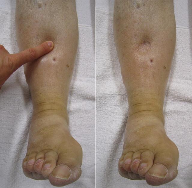
Palpation: Temperature, Turgor, Capillary Refill
Slide 20 lists four things to palpate: temperature, turgor and texture, capillary refill, peripheral pulses. Pulses get their own section next. For temperature the notes give a technique: use the dorsal aspect (the backs) of your hands and assess both extremities at the same time, comparing one with the other.
Why the backs of the hands, standard nursing content: the skin there is thinner and more sensitive to temperature than the palms.
| You feel | It indicates |
|---|---|
| One extremity cooler than the other | Arterial occlusion. “Evaluate for emergent nature of a condition.” Blood is not getting in. |
| One extremity warm, edematous and tender | DVT — in 30–40% of patients. “This also can be emergent.” Blood is not getting out. |
Turgor and texture share slide 22 with capillary refill. To test capillary refill, press on a nail bed until it blanches, release, and count the seconds until the colour returns. The slide only asks the question — “Normal Capillary Refill? (? Seconds)”. The instructor answers it.
The press-and-count technique is standard nursing content; the slide shows it in a photo without describing it.
Jeopardy gives “less than 2 seconds” as normal. The instructor said two to three, and what she said wins. If both appear, choose 2–3 seconds (under 3). Only a refill under 2 seconds is normal by either rule: exactly 2 seconds passes the instructor’s rule but not Jeopardy’s.
The Pulse Sites, by Name, Place and Purpose
Slide 23 names eight sites: temporal, carotid, brachial, radial, femoral, popliteal, posterior tibial, pedal. The notes add the technique — use the pads of two or three fingertips — and that the carotid and apical are covered with the heart — the apical with the heart sounds (Part 3), the carotid in the cardiac assessment (Part 5). The lecture adds the apical to the list of reasons, so it is here too. Slide 26 is five photos with no labels, and the instructor’s note is “Have students identify names of pulse sites”; the lecture asks over them, “Do you know the landmarks around each of these photos? Do you know the names of those landmarks?” — so the landmark is part of the answer.
| Pulse | Where to feel it | Used for (lecture) |
|---|---|---|
| Temporal | Over the temple, just in front of the upper ear† | When the radial is not accessible. “Not routinely used/assessed in adults” (slide 24 notes) |
| Carotid | Side of the neck, in the groove beside the trachea† — one side at a time | Cardiac arrest (the unresponsive adult) |
| Apical | 5th left ICS, midclavicular line — the apex, the PMI | Routinely in infants and children; to find a discrepancy with the radial — “and you should know this” — the pulse deficit = apical − radial (Jeopardy; Part 3); with some medications |
| Brachial | Inner upper arm, just above the elbow crease, beside the biceps tendon† | Blood pressure; cardiac arrest in infants |
| Radial | Thumb side of the inner wrist† | Routine — readily accessible |
| Femoral | The groin, just below the inguinal ligament† | Cardiac arrest and shock — Jeopardy: the alternative when the carotid cannot be felt |
| Popliteal | Behind the knee, deep in the hollow, knee slightly bent† | Circulation to the lower leg |
| Posterior tibial | Behind and below the medial malleolus — the inner ankle bone | Circulation to the foot |
| Dorsalis pedis (“pedal”) | The top (dorsum) of the foot, beside the tendon to the big toe† | Circulation to the foot |
† Standard anatomical landmark; the course documents name the site without describing it. Posterior tibial “below the medial malleolus” is Jeopardy’s, which also defines the medial malleolus: “the small prominent bone on the inner side of the ankle at the end of the tibia.”
The lecture wants you able to “pick out” pulse sites on a test. Start hides the names and asks for a pulse — click its dot. Before the next one appears, say why that site is used.
Drag sideways to see the whole figure →
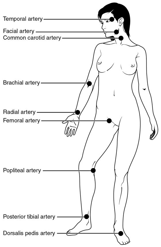
From the lab-manual table: use light palpation with the finger pads, and note rate, rhythm, equality, amplitude and elasticity; normal is regular, equal, +2, with soft, pliable arteries and no thrills. It also says to document pulse amplitudes on a stick figure — the same kind of figure the exam uses. The table lists an ulnar pulse too; the course deck does not. Its grading list also skips 2 — “0 = Absent, 1 = Weak, 3 = Full, 4 = Bounding” — while the same table gives normal as “+2”: answer with the deck and the lecture, 2+ is normal, documented 2+/4. “If indicated”, the table adds an Allen test (arterial flow to the hands) and an ankle–brachial index (flow to the legs, Part 7).
The Allen test, standard nursing content (Taylor uses it before a radial arterial blood gas): press on the radial and ulnar arteries while the patient makes a tight fist, have them open the blanched hand, release the ulnar — colour returning within a few seconds means the ulnar artery alone can supply the hand.
| Grade | Slide 25 |
|---|---|
| 0 | Absent |
| 1+ | Weak, thready, diminished |
| 2+ | Normal, expected — documented 2+/4 |
| 3+ | Increased, full |
| 4+ | Bounding |
Auscultation: Bruits and the Doppler
Slide 27 has two items. Bruit (carotid / aortic / femoral): the lecture — “we utilize the bell of our stethoscope to listen for bruits. And that’s a swishing sound.” And Doppler pulses if not palpable.
The recording of a real carotid bruit is in Part 3 (Bruits and Thrills), next to the murmurs it must not be confused with. In an adult, a normal carotid makes no swish; the lab-manual table notes a bruit “may be normal in children and with high-output states”.
| Step | What the course documents say |
|---|---|
| Palpate | One side at a time — “to ensure adequate cerebral blood flow” (cardiac deck). Pressing both at once can cut off the blood supply to the brain. Used in cardiac arrest. |
| Auscultate | With the bell, breath held. Normal (adult): no bruit. |
| Abnormal: a bruit | A swishing sound that means narrowing of the vessel (review sheet) or “a narrowing or disruption of blood flow” (Jeopardy) — turbulent flow. |
| Causes | A diseased artery — atherosclerotic plaque narrowing the lumen — or an enlarged thyroid gland pressing on the carotids: “An enlarged thyroid can cause turbulent blood flow in carotid arteries… if thyroid gland enlarged auscultate for bruit” (HEENT summary handout). |
The lab-manual cardiovascular table gives the technique: “Listen for bruits with bell of stethoscope. Have patient hold breath when auscultating for carotid bruits” — so breath sounds do not mask a soft bruit (Part 3 quotes the same line).
Standard nursing content: a bruit over a narrowed carotid is a stroke risk — the HEENT case client with a soft bilateral carotid bruit also had a blood pressure in the 180s.
The same lab-manual table pairs the carotid with the jugular vein: listen with the bell, breath held, for a venous hum. Normal is none, though a venous hum “may be normal” in children. To tell it from a murmur carried up from the heart, press on the jugular vein — a venous hum disappears.
Blood pressure — the lab-manual lines
The deck does not cover blood pressure; the lab-manual peripheral-vascular table lists it under auscultation:
| Line | What the table says |
|---|---|
| Where | Both arms; supine, sitting and standing. |
| Orthostatic drop | On standing, a fall in systolic of 10–15 mm Hg with a rise in pulse. Normal: no orthostatic drop. |
| Pulse pressure | Systolic minus diastolic — normally about one-third of the systolic (120/80 → 40). |
| Auscultatory gap | Palpate the brachial pulse, inflate until it disappears, then go 30 mm Hg higher before you listen — the same 30 as the vital-signs skill checklist. |
| Sounds down to “0” | Retake it and record Korotkoff 1, 4 (first diastolic) and 5 — all three. |
Two lab-manual figures are older than current practice: it calls a 10–15 mm Hg systolic fall orthostatic (current definitions use 20 systolic or 10 diastolic) and gives normal adult BP as under 140/90 (current guidelines call 130–139/80–89 stage 1 hypertension). If an item quotes the table, answer with the table.
The Doppler
When a pulse cannot be felt, it can often still be heard. The slide 27 notes, read aloud: the Doppler “can assess weak peripheral pulses. It magnifies pulsatile sounds from the heart and blood vessels as an arterial signal. The arterial signal is a rhythmic whooshing sound.” The technique, in the deck’s order:
| # | Step |
|---|---|
| 1 | Apply a drop of ultrasonic gel to the transducer |
| 2 | Place the transducer slightly angled over the artery |
| 3 | Turn on the volume and listen for the rhythmic whoosh |
Standard nursing content: the probe is usually held at about 45 degrees, pointing toward the heart, with light pressure (pressing hard can flatten the artery); once found, the spot is often marked with a pen so the next nurse can find it again. A pulse heard only by Doppler is commonly charted as “D” or “Doppler” rather than a number.
Labs: Arterial Risk, Venous Clot
| Side | Test | What it is for |
|---|---|---|
| Arterial disease | Lipid levels | Blood tests “to evaluate risks for PAD”. Targets from the cardiac deck: total <200, LDL <100, HDL >40, triglycerides <150 (Part 5). |
| HbA1c | “For diabetic patients” — long-term glucose control; diabetes is 4× the PAD risk. | |
| C-reactive protein and homocysteine | Further risk markers for PAD. | |
| Venous | D-dimer (blood test) | “D-dimer if DVT is suspected.” |
| Ultrasound venous Doppler study | Images the deep veins to look for the clot itself. The tidbits handout: a DVT is “best determined by venous duplex study (ultrasound)”. |
Standard nursing content: C-reactive protein is a marker of inflammation, and homocysteine is an amino acid whose high levels are linked to vessel damage and clotting. D-dimer is a fragment released when a clot breaks down; a normal D-dimer makes a DVT unlikely, but a raised one is not specific, so the ultrasound (venous duplex) is what confirms it.
The Seven P’s of Arterial Occlusion
The lecture: “So the seven P’s of arterial occlusion in limb. So this is a great way to remember.” They are what a limb looks and feels like when its artery is blocked — limb ischemia — and the review sheet points straight at them: “What are signs of Peripheral Arterial Occlusion/Disease? (See 7 P’s PPT from Peripheral Assessment).”
| # | P | What you find |
|---|---|---|
| 1 | Pain | In the limb below the blockage — rate it |
| 2 | Pallor (perfusion) | Pale skin — no blood arriving |
| 3 | Pulseless | Weak or absent pulse below the block — 0 or 1+ |
| 4 | Paresthesia (sensation) | Numbness, tingling, pins and needles |
| 5 | Polar / poikilothermic (coldness) | The limb is cold — it takes the temperature of the room |
| 6 | Paralysis | Cannot move it — muscle and nerve starved of oxygen |
| 7 | Perfusion | Capillary refill — delayed past 3 seconds |
The automatic transcript of the recorded lecture renders the list as “pain, power, pulselessness…”; the slide makes clear the word is pallor.
Standard nursing content: in acute limb ischemia, paresthesia and paralysis are late signs — the nerves and muscle are already being damaged, and the limb is in danger. Pain and pallor usually come first.
Clinical Judgement: Problems, Goals, Interventions
The last slide runs the findings through the clinical judgement model — hypothesis, generate solutions, take action, evaluate. The lecture: “we see that a patient has pale or cold extremities. So they might have altered tissue perfusion, arterial.”
| Hypothesis / problem | Cues |
|---|---|
| Altered tissue perfusion, arterial | Pale, cool extremities (“any of the 6 ps”); pain |
| Risk for peripheral neurovascular dysfunction | The same cues — a limb at risk of nerve and vessel damage |
| Activity intolerance | Pain and claudication with ambulation |
| Goals (evaluate against these) | Interventions (take action) |
|---|---|
| Peripheral pulses are strong and symmetrical | Monitor peripheral pulses every 4 hours |
| Capillary refill is <3 seconds | Assess and document edema with the scale every 4 hours |
| Peripheral edema is decreased | Evaluate pain on a 10-point scale |
| Patient senses sharp and dull accurately | Educate on risk factors |
| Patient verbalises a decrease in pain | Keep limbs warm; skid-free slippers |
| Through teach-back: understands the treatment regimen (exercise, medications, healthy behaviours) and the risk factors | Meticulous foot care once a day · ROM exercises to promote circulation · SCDs to promote venous return and reduce the risk of DVT |
That is the assessment. Part 7 takes the same findings and sorts them into the disorders the blueprint and the tidbits handout name — PAD, Raynaud’s, Buerger’s, venous thrombosis and DVT, venous insufficiency, varicose veins — with the arterial-against-venous comparison and the case of JH.
Arterial or Venous?
Part 6 taught you how to examine a leg. This part is about what the findings mean, and almost every question on it turns on one decision you make first: is the problem on the way in, or on the way out?
Arteries bring blood in. When they narrow, the tissue below the narrowing is starved — it is pale, cold, hairless, painful when it is asked to work, and it heals badly. Veins take blood out. When they or their valves fail, blood pools — the leg is swollen, warm, stained brown, and it aches after standing. Starved against flooded. Nearly every row of the table below follows from that one difference.
The course’s words for the two sides: the tidbits handout puts the arterial disorders under Peripheral Arterial Disease (PAD) and the venous ones under Peripheral Vascular Disease (PVD), and the peripheral-vascular preparation sheet tells you to “review and understand difference between PAD and PVD.” The blueprint’s heading for this whole area is “Refer to handouts and case summaries”, so the handout and case JH are the backbone of this part.
| Arterial — PAD | Venous — insufficiency, DVT, varicose veins | |
|---|---|---|
| What fails | Blood cannot get in: atherosclerosis narrows the artery and tissue below the narrowing is damaged (tidbits) | Blood cannot get out: incompetent valves and stretched veins let it pool — venous stasis (tidbits) |
| Pain, and when | Intermittent claudication — cramping in the calf, thigh or buttock on walking, gone with rest. Later, rest pain at night, relieved by hanging the leg down (tidbits) | Dull ache, fullness in the legs after standing (varicose veins); DVT: calf or groin tenderness (tidbits). Eased by raising the leg |
| Pulses | Decreased or absent (tidbits) | Usually present† |
| Colour | Pallor when the legs are up, rubor (red) when dependent; cold grey-blue (tidbits) | Brownish discoloration from the ankles up to the calf (tidbits); DVT: red (erythema, deck slide 18) |
| Temperature | Cool or cold. One limb cooler than the other = arterial occlusion (deck slide 21) | Warm. Warm, edematous and tender = DVT (deck slide 21) |
| Hair and skin | Hair loss; dry, scaly skin (tidbits); thin and shiny (case JH) | Hair usually present†; stasis dermatitis — the brown staining (tidbits) |
| Nails | Thickened toenails (tidbits, case JH) | Usually normal† |
| Edema | Usually none (case JH: “No edema”) — but swelling would worsen arterial flow (tidbits) | The hallmark. “Edema can indicate venous abnormalities” (deck slide 13) |
| Ulcer | On or between the toes, the top of the foot, the lateral ankle — painful (tidbits, Jeopardy). Deep, “punched-out”, pale base† | Venous stasis ulcer around the ankle, classically the inner (medial) ankle†; can lead to cellulitis (tidbits). Shallow, irregular, moist† |
| Positioning | May raise the legs while sitting in a chair, but never above the heart; in severe PAD, dangle the legs over the bed edge (tidbits) | Elevate — above the heart in bed, and 10–20 minutes every few hours (tidbits) |
| Warmth | Avoid cold; insulated socks and shoes. No heating pad directly on the legs (tidbits) | Phlebitis and DVT: warm, moist compresses as ordered, “not too hot” (tidbits) |
| Compression | Tight wraps endanger arterial flow — the handout warns an Unna boot (below) that is too tight can cause arterial occlusion | Stockings are central — on before getting out of bed, perhaps for life (tidbits) |
| Exercise | Walk to the point of claudication, rest, then walk a little farther — builds collateral circulation (tidbits) | Progressive ambulation; avoid prolonged sitting or standing (tidbits) |
| Both | Avoid crossing the legs and constrictive clothing; stop smoking; take medications as prescribed; check the skin of the legs and report changes (tidbits, on both sides of the handout). | |
Drag sideways to see the whole figure →
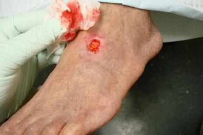
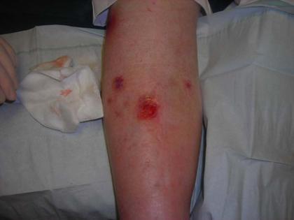
Peripheral Arterial Disease
The handout’s definition: a chronic disorder in which partial or total arterial occlusion deprives the lower extremities of oxygen and nutrients, and tissue damage occurs below the level of the occlusion. Atherosclerosis is the most common cause — narrowing from plaque, the same process as coronary artery disease, and the handout says its risk factors are “very much like in CAD.”
| Finding | Why it happens |
|---|---|
| Intermittent claudication | “Pain in muscles resulting from inadequate blood supply” — walking raises the muscles’ demand for oxygen and the narrowed artery cannot meet it. Rest lowers the demand, so the pain stops. |
| Rest pain | Numbness, burning, aching in the distal leg that wakes the patient at night, relieved by a dependent position. Now the supply fails even at rest; lying flat removes gravity’s help, hanging the leg restores it. |
| Discomfort in the lower back or buttocks | The narrowing is higher up, in the arteries feeding the hips. |
| Hair loss; dry, scaly skin | Hair follicles and skin need a steady blood supply. The review sheet: “loss of hair on legs due to decreased circulation/oxygen.” |
| Thickened toenails | Chronic poor nourishment of the nail bed. |
| Cold, grey-blue skin | Too little warm arterial blood arriving. The review sheet says “cold, clammy skin on legs.” (“Clammy” is the review sheet’s word; the handout describes PAD skin as dry and scaly. If an option offers “cold, clammy skin” for arterial insufficiency, it is the review sheet’s answer.) |
| Pallor up, rubor down | Raised, the foot loses the little pressure it had and blanches; hanging, blood pools in widened small vessels and it turns red. |
| Decreased or absent pulses | Less blood is getting past the narrowing. Grade them and, if you cannot feel them, Doppler them (Part 6). |
| Arterial ulcers | On or between the toes, upper aspect of the foot — painful. Starved tissue breaks down and cannot heal. |
| Low leg blood pressures | BP at the thigh, calf and ankle is lower than the brachial. Normally leg pressures are higher than arm pressures, so a lower one means the leg is downstream of a narrowing. |
The leg-to-arm pressure comparison is what the ankle–brachial index measures (ankle systolic ÷ brachial systolic; about 1.0 or higher is normal, under about 0.9 suggests PAD). The lab-manual table mentions the index; the number is standard nursing content.
| Group | What the nurse does or teaches |
|---|---|
| Assess | Pain; colour, motion, sensation and pulses of the extremities — in bold: “Know the 6 Ps of assessment from the power point slide” (the P’s are in Part 6); BP measurements; signs of ulcer formation or gangrene. |
| Exercise | An individualised exercise program, started slowly and increased gradually. Prescribed exercise develops collateral circulation. Walk until the point of claudication, stop to rest, then walk a little farther. |
| Protect the flow | No leg crossing, no restrictive clothing. Avoid cold — it causes vasoconstriction. Insulated socks and shoes — warmth promotes vasodilation. |
| Protect the skin | Do not apply heat (a heating pad) directly to the legs — “possible neuropathy could lead to burns.” Teach the patient to assess the skin of the lower legs and report changes. |
| Medication | Emphasise compliance with the medication regimen. |
| Position | Elevate the legs while resting in a chair — not higher than the heart. Severe PAD: sleep with the leg dangled over the edge of the bed or sit with the legs dependent, for comfort and perfusion to the feet. |
Acute Occlusion, Procedures and Embolectomy
The handout draws a line you should hold onto: everything above is chronic PAD, built up over months or years. A patient with an acute occlusion from an embolus — a clot that has travelled and lodged — is an emergency and “may require an emergency procedure: catheter embolectomy, thrombolytic or surgical embolectomy, depending on severity.”
The acute picture is the P’s of arterial occlusion / limb ischemia (Part 6) and the deck’s one red line: pain, numbness and tingling, coolness or colour change — “urgent/ possible total arterial occlusion.” Jeopardy lists the findings that “need immediate intervention”: absent or diminished pulses, delayed capillary refill, pallor, and numbness and tingling.
| Chronic PAD | Acute arterial occlusion | |
|---|---|---|
| Onset | Gradual — “worsening over the past few months” (case JH) | Sudden |
| Pain | Claudication with walking, eases with rest | Pain that does not ease, with numbness and tingling |
| Limb | Cool, pale on elevation, hairless, thick nails | One limb suddenly colder and paler than the other, pulseless; paralysis late |
| Nurse | Teach, exercise, protect, follow up | Notify the provider immediately; prepare for embolectomy or a clot-buster |
| Procedure | What it is |
|---|---|
| PTCA, with or without stent | Percutaneous transluminal angioplasty: a catheter into the artery, inflate the balloon, and place a stent to hold it open. (The handout writes PTCA, the coronary abbreviation; on a leg artery it is usually called PTA.) |
| Laser-assisted angioplasty | The same idea, using a laser to clear the narrowing. |
| Bypass surgery | A graft routes blood around the blockage — aorto-femoral or femoral-popliteal bypass. |
| Surgical embolectomy | For acute obstruction: removal of an embolus from an artery using a catheter; may need a patch graft on the artery. |
| Before | After |
|---|---|
| Baseline vascular assessment — you need it to compare against afterwards. | Neurovascular assessment — the 6 P’s and motor function — plus neuro, cardiac and respiratory. Assess for re-occlusion. |
| Anticoagulants as ordered — heparin titrated to a therapeutic PTT. | O2 and pulse oximetry as ordered. Semi-Fowler’s position. |
| Thrombolytics (“clot buster”) as ordered — risk of bleeding; know there are contraindications. | Reperfusion complications: muscle swelling → compartment syndrome (pain) → fasciotomy to release it. The artery may also spasm — vasodilators. |
| Keep the extremity dependent to enhance perfusion. | Incision: bleeding, hematoma, infection. Labs: CBC (WBC for infection, Hgb/Hct for bleeding), electrolytes, clotting studies. |
| Anticoagulants and antiplatelets as prescribed; teach anticoagulant therapy, signs of infection and edema; avoid long sitting, crossed legs, restrictive clothing; how to wear, remove and reapply anti-embolism stockings. |
The handout also says, of every vascular procedure, that the peri-operative basics always apply: control pain, prevent pneumonia, prevent DVT, prevent wound infection.
Raynaud’s and Buerger’s
Both appear on the deck’s common-disorders slide and in the handout, and both affect the small arteries of the fingers and toes. The difference is spasm against inflammation: Raynaud’s vessels clamp down and then let go; Buerger’s vessels are inflamed and occluded.
| Raynaud’s disease | Buerger’s disease (thromboangiitis obliterans) | |
|---|---|---|
| What | Vasospasm of the arterioles and arteries of the upper and lower extremities → constriction of skin vessels | Inflammation — occlusive disease of the medium and small arteries and veins |
| Where | Fingers, toes, ears and cheeks | The distal parts of the arms and legs |
| Triggers / pattern | Intermittent attacks with cold or stress | Constant and progressive† |
| Findings | Blanching, then cyanosis during the spasm; redness when it is relieved. Numbness, tingling, swelling, cold | Intermittent claudication; ischemic pain worse at night; cool, numb, tingling; decreased distal pulses; cool and red when dependent; ulcers |
| Nursing | Monitor pulses; vasodilators as prescribed and teach them; identify and avoid triggers; smoking cessation; warm clothing, socks and gloves in cold weather; avoid injury to hands and feet | Stop smoking (listed first); monitor pulses; avoid injury to the extremities; vasodilators as prescribed and teach them |
Standard nursing content, not in the handout: Buerger’s disease is very strongly tied to tobacco use, typically in younger adult smokers, and stopping tobacco completely is the one treatment that halts it — which is why the handout lists smoking cessation first.
Assessing Risk and Preventing Arterial Disease
The blueprint asks for “assessing risk of arterial disease” and “prevention of arterial disease/ risk factors”. The first is a set of questions you ask (deck slide 14); the second is what you can change (deck slide 15). The handout says the risk factors are “very much like in CAD”, and the deck’s notes say the same: “same as risk factors for CAD that can lead to MI.”
Ask about (slide 14) — a personal or family history of:
| Ask about | Why, from the deck’s notes († standard nursing content) |
|---|---|
| Heart and circulation problems | Clots, ulcers, numbness, edema in the arms or legs. “CV disease = strong hereditary link.” |
| Smoking | “One of the most devastating” risk factors; stopping “can significantly delay the progression of atherosclerosis.” |
| Diabetes | Patients with diabetes are 4× more likely to develop lower-extremity PAD. A family history of diabetes raises personal risk. |
| Sedentary lifestyle; exercise | Inactivity is one of the three most modifiable factors. |
| Sitting or standing for long periods | Occupation and habits — this one matters on the venous side too. |
| Hypertension | Damages the artery lining.† |
| Medications | Oral contraceptives, estrogen. |
| Lipids; diet | High LDL, high triglycerides and low HDL are associated with atherosclerosis; a high-fat diet. |
| Recent trauma to an extremity | “Thromboembolism can develop as a result of blood vessel trauma.” |
| Modifiable — teach to these | Non-modifiable — assess, cannot change |
|---|---|
| Smoking — the most devastating | Genetics and family history of heart disease |
| High cholesterol; diet high in saturated fat | Age — PAD “increases dramatically in the seventh and eighth decades” (slide 12) |
| Sedentary lifestyle | Gender |
| Hypertension | Ethnicity |
| Diabetes (its control); obesity, especially around the middle | (Jeopardy’s list of non-modifiable atherosclerosis risk factors, exactly) |
The deck’s notes single out smoking, high-fat diet and limited activity as “the most modifiable.” The blood tests that measure arterial risk (slide 28): lipid levels, HbA1c for diabetic patients, and C-reactive protein and homocysteine. And slide 12: the American Heart Association says prevention begins in childhood — atherosclerosis already shows in young people with familial hypercholesterolaemia, so teach the risk factors early.
Venous Thrombosis
A thrombus can come with inflammation, and inflammation can come with a thrombus: the handout says the inflammation thickens the vein wall and leads to embolisation — the clot breaking loose. That is the whole danger of the venous side: a leg clot becomes a lung clot.
| Term | Meaning |
|---|---|
| Thrombophlebitis | Thrombus with inflammation |
| Phlebothrombosis | Thrombus without inflammation |
| Phlebitis | Vein inflammation associated with invasive procedures — IV lines |
| Deep vein thrombophlebitis (DVT) | In a deep vein — more serious than a superficial one because of the increased risk of pulmonary embolism (PE) |
| Risk factor | Examples from the handout |
|---|---|
| Venous stasis | Varicose veins, immobility, heart failure |
| Hypercoagulability | Hypercoagulability disorders; pregnancy; oral contraceptives; certain malignancies |
| Injury to the vein wall | IV catheters, IV medications (irritants to the wall); fractures or other injuries to the legs or pelvis |
| Surgery | Orthopaedic and abdominal surgeries carry the greatest post-operative risk |
The handout lists eight items; grouping them under stasis, clotting and vessel-wall injury is the textbook’s Virchow’s triad (standard nursing content). If a stem asks why a post-operative orthopaedic patient is at risk, all three apply: immobile (stasis), surgical trauma (wall injury), and the body’s clotting response to surgery.
| Phlebitis | Deep vein thrombophlebitis (DVT) | |
|---|---|---|
| Find | Warm, red area radiating up the vein; pain, swelling, soreness | Calf or groin tenderness / pain, with or without swelling; warm skin, tender to touch. Homans’ sign is not a reliable indicator. Best confirmed by venous duplex ultrasound; D-dimer if suspected (deck slide 28) |
| Do | Warm moist soaks as ordered to dilate the vein — not too hot; assess for tissue necrosis and infection | See the list below |
| Group | Intervention |
|---|---|
| Do not dislodge it | Bed rest as ordered. Avoid a pillow under the knee. Do not massage the extremity. |
| Help it drain | Elevate the extremity as ordered; anti-embolism stockings as ordered (teach how to apply and remove); warm, moist compresses as ordered. |
| Watch it | Monitor for warmth and edema; measure the circumference of the extremity. |
| Medicate | Thrombolytic, heparin, Coumadin as ordered (table below); analgesics; diuretics as ordered for swelling of the leg. |
| Teach | Avoid prolonged sitting or standing, constrictive clothing, crossing the legs; elevate the legs every few hours; a progressive ambulation program; stop smoking. |
| Drug | What it does | Lab | Watch and teach |
|---|---|---|---|
| Thrombolytic (“clot buster”) | Dissolves an existing clot | — | Bleeding; there are contraindications — know they exist |
| Heparin (IV or SQ) | Stops the clot getting larger and new ones forming — it does not dissolve it | PTT | Follow the heparin protocol; bleeding; heparin-induced thrombocytopenia (HIT) — a fall in platelets |
| Coumadin (warfarin), by mouth | Longer-term prevention of clots | PT / INR | Teach: diet, medic-alert bracelet, blood drawn at the prescribed interval, compliance, signs of bleeding, caution with injury |
| Lovenox (SQ); aspirin, Plavix | Anticoagulant; antiplatelets | — | Listed after embolectomy — “become comfortable with” them |
Pairing hook (standard): heParin → PTT (P with P…T…T), warfarin/Coumadin → PT/INR (“War” and “PT” are the shorter pair). The handout’s “Diet” means, in standard teaching, keeping vitamin K foods (leafy greens) consistent rather than cutting them out, because vitamin K counteracts warfarin.
The IVC filter
An inferior vena cava filter is a wire, umbrella-shaped filter placed in the inferior vena cava to trap emboli and stop them reaching the lungs (tidbits). The deck’s vein slide points to the IVC as the place it sits. It catches clots; it does not dissolve them or stop new ones forming in the leg.
| Before | After |
|---|---|
| Discontinue anticoagulants for the procedure; teach what to expect. | Vital signs; insertion site for bleeding, hematoma, infection; retroperitoneal (internal) hemorrhage — back pain, changing vital signs. |
| System assessment (neuro, respiratory, renal, cardiac); O2 and pulse oximetry; semi-Fowler’s — avoid hip flexion; progressive activity. | |
| Resume anticoagulants and monitor the labs; assess peripheral edema; maintain anti-embolism stockings. Teach: elevate the legs while sitting, avoid long sitting and crossed legs, how to remove and reapply the stockings, progressive ambulation. |
The recorded lecture describes the IVC filter as something that “actually prevents clots”. The handout’s wording is the precise one: it prevents clots moving to the lungs. If an option says a filter treats or dissolves the DVT itself, it is wrong.
Preventing Venous Emboli — The Checklist
Three course documents ask the same question three ways: the blueprint line, the review sheet’s “What interventions are useful in prevention of VTE (Venous Thrombus Embolism)?”, and the deck’s capitals, “HOW CAN WE HELP TO ENHANCE VENOUS RETURN IN OUR HOSPITALIZED PATIENTS?” The answer to all three is the same idea: keep the blood moving out of the legs, because blood that sits still clots.
| Intervention | How it keeps blood moving | |
|---|---|---|
| ☐ | Encourage ambulation — early, progressive | Walking works the calf muscle pump, which veins depend on because they have no muscular wall of their own (deck slide 10). Review sheet; tidbits. |
| ☐ | SCDs (sequential compression devices) | “They simulate that calf muscle pump for hospitalized or bed-bound patients, particularly post-surgery” — said in the lecture. Review sheet; deck slide 31. |
| ☐ | Anti-embolism stockings as ordered | Squeeze the superficial veins so blood moves into the deep veins and up. Teach how to put them on, remove and reapply them. |
| ☐ | Elevate the legs while sitting, and every few hours | Gravity helps return. Review sheet; tidbits (DVT, IVC filter). |
| ☐ | No crossing the legs; no constrictive clothing | Both compress veins and slow return. Tidbits, every venous section. |
| ☐ | Avoid prolonged sitting or standing | Long stillness = stasis. Tidbits; the review sheet’s PVD list (“Avoid standing long periods?”). |
| ☐ | Range-of-motion exercises | Active or passive ROM moves the calf when walking is not possible. Deck slides 2 and 31. |
| ☐ | Assess dorsalis pedis and posterior tibial pulses regularly | Not a preventer in itself — it is how you catch a change early. The review sheet lists it; the deck’s notes say monitor pulses every 4 hours and grade edema every 4 hours. |
| ☐ | Stop smoking | Tidbits, DVT interventions. |
| ☐ | Anticoagulants as ordered | Heparin or Lovenox for patients at high risk — collaborative, not a nursing decision.† |
† Standard nursing content: the handout names heparin and Coumadin for an existing DVT and lists Lovenox only after embolectomy; it does not give a prevention dose for high-risk patients.
Standard nursing content: dehydration thickens the blood and raises VTE risk, so adequate hydration is part of prevention. Fluid restriction belongs to heart failure, not to vascular disease.
Venous Insufficiency and Varicose Veins
Venous insufficiency comes from prolonged venous hypertension — high pressure in the veins stretches them and damages the valves. With incompetent valves, blood pools: venous stasis and edema, which lead to venous stasis ulcers, swelling and cellulitis. Assessment: dermatitis from venous stasis — brownish discoloration along the ankles, extending up to the calf; edema; ulcers. The treatment focus is to decrease edema and promote venous return, and once an ulcer exists, to heal it and prevent it coming back.
| Teach | Detail |
|---|---|
| Stockings | Elastic or compression stockings during the day and evening; apply before getting out of bed; may need them for the rest of their life. |
| Elevation | 10–20 minutes every few hours; above the heart while in bed. Varicose veins: “as much as possible.” |
| Avoid | Prolonged sitting or standing; crossing the legs when seated; constrictive clothing. |
| Home compression | A home compression system — the handout compares it to the hospital’s device. With an ulcer, keep a dressing over it so the device never touches the ulcer directly. |
| Wound care | As ordered; assess the patient’s ability to care for the wound at home; occlusive dressings such as hydrocolloid; Unna boot (below). |
| Medications | Antibiotics if cellulitis develops; topical agents to remove dead tissue (e.g. Silvadene cream) — not on healthy tissue. |
The handout says the home system is “like ICD in the hospital”. In context that is almost certainly a typing slip for SCD — the sequential compression device the lecture discusses.
| From the handout | |
|---|---|
| What | “Distended, protruding veins that appear darkened and tortuous”; the vein walls weaken and distend. |
| Assess | Dull aching pain after standing; a feeling of fullness in the legs; ankle edema. |
| Valve test | The lab-manual table: “If varicosities present, check venous valve competence with Trendelenburg test or manual compression test.” (Standard: manual compression — press the dilated vein low down and feel 15–20 cm higher; an impulse felt there means the valves between are incompetent. Trendelenburg test — raise the leg to empty the veins, compress the upper thigh, stand the patient; veins that fill fast from above once the pressure comes off have incompetent valves. Not the Trendelenburg position.) |
| Nursing | Anti-embolism stockings (emphasise them); elevate the legs as much as possible; no crossed legs or constrictive clothing; prepare for procedures. |
| Sclerotherapy | A solution injected into the vein, then a pressure dressing; a follow-up incision 2–3 weeks later removes trapped blood. |
| Laser therapy | A laser fibre heats and closes the main vessel feeding the varicosity. |
| Vein stripping | Removal of large or clustered varicose veins, or when other treatments fail. Before: mark the vein; baseline peripheral vascular assessment. After: ace wraps; monitor bleeding, infection, mobility, nerve damage, pulses; elevate the legs; elastic stockings once the bandage comes off; range-of-motion exercises. |
The Rest of the Deck’s List
The deck’s common-disorders slide names ten conditions, and the lecture tells you plainly to look up any you do not know. Most are covered above. The three the handout does not cover are below, in one line each.
| Disorder | In one line |
|---|---|
| Peripheral arterial disease | Partial or total occlusion; the P’s. Above. |
| Claudication | Pain with walking, relieved by rest — see the trap on the slide’s wording above. |
| Buerger’s disease | “Occlusive disease of small arteries.” Above. |
| Abdominal aortic aneurysm (AAA) | A ballooning of the aorta in the abdomen where its wall has weakened.† Sometimes felt as a pulsating mass; a bruit over the aorta is one of the deck’s auscultation sites (slide 27). Do not palpate deeply over a known or suspected one.† |
| Raynaud phenomenon and disease | Vasospasm with cold or stress — white, blue, red. Above. |
| Chronic venous insufficiency | Damaged valves, pooling, brown staining, ulcers. Above. |
| Deep vein thrombosis | Clot in a deep vein; PE risk. Above. |
| Thrombophlebitis | Clot with inflammation. Above. |
| Neuropathy | Nerve damage — numbness, tingling, burning, loss of sensation in the feet. Diabetes is a common cause (Jeopardy). It is why the PAD patient gets no heating pad and daily foot checks: they may not feel a burn or a wound. |
| Lymphedema | Swelling from lymph that cannot drain. The deck’s lymphatic slide: the lymph system maintains fluid and protein balance, and “overload — lymph edema.” Typically after lymph nodes are removed or damaged (for example, breast-cancer surgery).† |
† Standard nursing content; the deck names these without defining them.
Case JH, Worked Through
JH is a 55-year-old married African American man admitted with a history of CAD, hypertension and diabetes. Chief complaint: “My left leg hurts and I have two ulcers on my toe that will not heal.” The case sheet walks you through the clinical judgement model — the same model as the deck’s slide 31 — one heading at a time. Here it is filled in.
| Subjective | Objective |
|---|---|
| Left-leg pain when walking, sooner on inclines and stairs, gone with rest | Bilateral DP and PT pulses absent in the left leg (see the note below) |
| “Pressure and tightening, sometimes sharp and cramp-like”, in the calf and thigh muscles | Feet pale when elevated, cool to touch |
| Occasional for a month, now daily; walking harder | Thin, shiny skin; patchy hair loss on the legs; thickened toenails |
| Two toe ulcers that “will not heal” | Two small open ulcers on the left great toe |
| Uses no pain medication | No edema, no varicosities — the venous side is clear |
| Home BP 150–160 / 90; fasting sugar 140–150 | BP 150/96, HR 98, RR 16, afebrile |
| “Hard to eat healthy”, cheats on his diet; “too busy” to exercise; dinner and TV | 5 ft 9 in, 220 lb — obese abdomen |
| Leg pain is stopping him going to the movies and dinner with friends | Alert and oriented ×3; lungs clear; S1 S2 normal; urine adequate |
Arithmetic you can check: 220 lb is about 100 kg and 5 ft 9 in is about 1.75 m, so his BMI is about 32.5 — obese (30 and over), standard content. A normal fasting glucose is under 100 mg/dL, so 140–150 is well above it.
| Modifiable | Non-modifiable |
|---|---|
| Diabetes — ten years, poorly controlled (4× PAD risk) | Age 55, male |
| Hypertension — above target on medication | Ethnicity — African American |
| Obesity; a diet he struggles to follow | Family history: father died of an MI at 57; mother had a stroke at 62 and type 2 diabetes |
| Sedentary — no exercise; long hours and frequent travel as a salesman (long sitting) | Existing CAD — atherosclerosis already present in one bed |
Note what is not there: the case never mentions smoking. Do not add it. A nurse assesses it — it is the first question on slide 14 — but you do not teach cessation to a man who has not been shown to smoke.
| Step | For JH |
|---|---|
| Analyse cues — arterial or venous? | Arterial. Claudication relieved by rest, absent pulses, pallor on elevation, coolness, hair loss, shiny skin, thick nails, a painful ulcer on the toe — and no edema, no varicosities. Chronic, not acute: the pain has built over months and still goes away with rest. |
| Hypotheses — actual | Altered tissue perfusion, arterial (left leg) · Activity intolerance — pain and claudication with walking (both exactly as deck slide 31 words them) · Impaired skin integrity — two open toe ulcers · Pain · Ineffective health management — BP and glucose above target, diet, no exercise |
| Hypotheses — risk for | Risk for peripheral neurovascular dysfunction (deck slide 31) · Risk for infection — open wounds, poor flow and diabetes · Risk for injury — ten years of diabetes, so possible neuropathy |
| Prioritise | 1. Altered tissue perfusion, arterial — it is the cause of almost everything else, and if it worsens to an acute occlusion the leg is at stake. 2. Impaired skin integrity / risk for infection — non-healing ulcers on a poorly perfused diabetic foot are how infection and gangrene start. Activity intolerance and pain follow from the first and improve with it. |
| Generate solutions — goals | From the deck’s slide 31 notes: peripheral pulses strong and symmetrical; capillary refill under 3 seconds; patient senses sharp and dull accurately; through teach-back, states the treatment plan — exercise, medications, healthy behaviours — and the risk factors; reports less pain. For the ulcers: no signs of infection, wound shrinking. |
| Take action | Assess: pulses every 4 hours (Doppler if not palpable), the P’s, pain on a 0–10 scale, the ulcers for infection or gangrene, BP. Teach: walk to the point of claudication, rest, walk a little farther; legs not above the heart; keep the feet warm with socks and shoes but no heating pad; meticulous daily foot care and skin checks; no crossed legs or tight clothing; take medications as prescribed. Collaborate: wound care as ordered; dietitian; provider about BP and glucose control. Start with what JH is willing to work on — getting back to the movies with his friends is a goal walking serves. |
| Evaluate | Were the goals met? Pulses (by Doppler at least), capillary refill, pain scores, how far he walks before claudication — the distance should grow — ulcer appearance, and his teach-back. Any sudden pain at rest, colder or paler foot, numbness or weakness is no longer evaluation — it is an acute occlusion; notify the provider now. |
Standard nursing content for the diabetic, poorly perfused foot that the case does not spell out: never walk barefoot, check between the toes, keep the skin moisturised but not between the toes, and have a professional cut the thickened nails.
What the Blueprint Asks
The sleep block is six questions, and all three blueprint lines end in the same two words: “PP slides”. That means the Rest & Sleep lab deck, not the textbook chapter. When the deck and Taylor disagree in this part, the deck wins and Taylor’s version sits underneath in grey.
| Blueprint line | Deck slides | In this part |
|---|---|---|
| “Nursing sleep interventions” | 25 (with 24 and 26) | Helping Patients Sleep — the ten interventions, the bedtime snack, narcolepsy teaching |
| “REM and NREM characteristics” | 8, 9, 10 | NREM and REM — the drawn arrows, the stage-by-stage table |
| “Understand normal sleep cycles and chemicals present” | 6, 7, 11, 24 | What Runs Sleep (melatonin, the hypothalamus, circadian rhythm) and The Sleep Cycle (the night, drawn) |
“Chemicals present”, all in one line. The deck has three: melatonin regulates the sleep–wake cycle (slide 6) and “helps to induce sleep”, and carbohydrates stimulate it (slide 24, red); protein wakens the brain (slide 24, red); and short sleep lowers leptin, which raises appetite (slide 21) — the only one the deck itself calls a “chemical change”, and the reason obesity heads slide 5’s red list. Melatonin is in What Runs Sleep; leptin is in the next section.
Rest, Sleep and What Losing It Costs
The deck separates two words that people use interchangeably. Rest is a decreased state of activity that leaves you feeling refreshed — you can rest wide awake. Sleep is a state of rest with an altered state of consciousness and relative inactivity, whose job is to restore mental and physical function. Sleep is not one flat state: it is a complex rhythmic state, a progression of repeated cycles, each with different phases of body and brain activity. That last phrase is the whole of the NREM–REM section to come.
Slide 4: “Adequate amounts of rest & sleep promote health / Critical for physical/mental/ & emotional wellbeing.”
Slide 5: “Chronic sleep loss can increase risks: obesity, depression, hypertension, diabetes mellitus, heart attack, stroke.”
Learn the six risks as a set — a “which of these is not a risk of chronic sleep loss” item is the obvious question to build from a red list.
| Kind of effect | What you see (slides 5 and 21) |
|---|---|
| Short-term — sleep deprivation | Inability to concentrate, poor judgement, moodiness, irritability, increased risk for accidents. |
| Physical and psychological | Low energy level, decreased self-worth, changes in facial and behavioural characteristics. |
| Chemical | Decreased leptin → increased appetite. That is the mechanism behind obesity on slide 5, and why short sleep in childhood raises the risk of obesity. |
| Long-term — chronic loss (red) | Obesity, depression, hypertension, diabetes, heart attack, stroke. |
| Physical data suggesting a sleep problem | Snoring; nocturnal myoclonus — muscle jerking in sleep. |
Taylor adds that REM is believed to play a role in learning, memory and adaptation, and that sleep and wake states carry distinct hormonal patterns that influence metabolism and glucose homeostasis — one way to make sense of diabetes and obesity on the red list. The deck gives each kind of sleep its job in the stage table further down.
What Runs Sleep
Slide 6 names three brain structures and one hormone. Learn them as a short chain: two systems in the brainstem switch sleep on and off in cycles, the hypothalamus is the control centre, and melatonin is the chemical messenger that tells the body it is time to sleep.
| Structure or chemical | What the deck says it does |
|---|---|
| Reticular activating system (RAS) | In the brainstem. Facilitates reflex and voluntary movement and controls the cortical activity of alertness. During sleep it receives few stimuli from the cerebral cortex and the periphery — so stimulation (noise, pain, light) is what wakes you. |
| Bulbar synchronizing region | Also in the brainstem. Works with the RAS to control the cyclic nature of sleep. |
| Hypothalamus | Controls several involuntary activities; it is the control centre for sleeping and waking. Injury to the hypothalamus → sleeping for abnormally long periods. |
| Melatonin | A hormone thought to regulate the sleep–wake cycle, and possibly the circadian rhythm. This is the “chemical present” the blueprint means. |
“Protein wakens the brain—good to have in the morning.
Melatonin helps to induce sleep—carbohydrates stimulate melatonin.”
So the chemistry and the snack are one fact: a carbohydrate at bedtime → more melatonin → sleep comes more easily. A protein at bedtime does the opposite. The bedtime-snack question is answered from this slide — see Helping Patients Sleep.
The circadian rhythm (slide 7)
A circadian rhythm completes a full cycle every 24 hours. Circadian synchronization is when your sleep–wake pattern follows your inner biological clock. That clock is influenced by light and dark — but the deck is careful to add, “not primary control” — and by occupational demands and social pressures. Desynchronization is what happens when the sleep–wake pattern is frequently altered: rotating shifts, jet lag, a hospital stay.
Standard physiology, beyond the deck and in grey because the blueprint says “PP slides”: Taylor lists the neurotransmitters involved — norepinephrine, acetylcholine, dopamine, serotonin and histamine with excitation, GABA with inhibition — and explains caffeine as blocking adenosine, a compound that builds up while you are awake and causes drowsiness. Taylor also explains the carbohydrate effect through the amino acid L-tryptophan (carbohydrates make it more available to the brain). If a question offers melatonin among these, the deck’s answer is melatonin.
NREM and REM
There are two kinds of sleep. NREM (non-rapid eye movement) is most of the night — 75–80% — and is split into four stages that get deeper as they go. REM (rapid eye movement) is the other 20–25%: the dreaming sleep. The cleanest way to keep them apart is the one the instructor drew on slide 8 — the vital signs go in opposite directions.
| NREM | REM | |
|---|---|---|
| Share of sleep | 75–80% | 20–25% |
| Nervous system | Parasympathetic (PNS) dominates | (the slide does not say for REM) |
| Temperature, HR, RR, BP, metabolic rate | ↓ all five | ↑ all five — and they vary (slide 10: “varying VS”) |
| Skeletal muscle tone & reflexes | Relaxed | ↓ — the body is nearly still while the brain is busy |
| Waking the person | Easy in I and II; hard in III; very hard in IV | More difficult to arouse than NREM |
| Dreams | — | Vivid dreaming; woken in REM, the person remembers dreams |
| What it restores | The body — stage IV: physiologic rest, repair and renewal of tissue | The mind — cognitive restoration; “believed to be essential for mental & emotional stability” |
| If deprived | — | REM rebound: “REM will increase in successive nights” |
REM: the brain lights up and the body goes limp — vital signs ↑ and irregular, muscle tone ↓, vivid dreams.
The four NREM stages, and REM
Slide 9 lays the four stages out as a two-by-two grid, and the text runs in a confusing order if you read it as a list — Stage 1 top left, Stage 3 top right, Stage 2 bottom left, Stage 4 bottom right. Read by box, this is what each stage says:
| Stage | Share | Length | Vital signs | Waking | What else |
|---|---|---|---|---|---|
| NREM I | ~5% | Only a few minutes | VS & metabolism begin to decrease | Awakens easily | Very light sleep; feels relaxed and drowsy |
| NREM II | ~50% | 10–20 min | VS & metabolism continue to slow | Slightly more stimulation needed | “Deeper sleep”; increased relaxation |
| NREM III | ~10% | 15–30 min | Continue to decrease but remain regular | Difficult | Initial stage of deep sleep; relaxation with little movement |
| NREM IV | ~10% | 15–30 min | VS low | Very difficult | Delta sleep — deepest sleep. Physiologic rest and restoration; repair and renewal of tissue. Enuresis, sleepwalking, sleep talking happen here. |
| REM | 20–25% | Average 20 min, longer each cycle | Varying (↑) | Very difficult | First REM about 90 minutes after falling asleep. Vivid dreaming; cognitive restoration. |
Taylor 10e Box 35-1 adds detail the deck leaves out. Stage I can include involuntary muscle jerking that wakes the person; stage II is 50–55%. In REM the eyes dart, small facial muscles twitch, large muscles are immobile, resembling paralysis, respirations are irregular, sometimes with apnea, the pulse is rapid or irregular, gastric secretions increase, and the EEG is active. Taylor describes the arousal threshold as greatest in stage IV.
The Sleep Cycle
Slide 11 is four sentences, and together they describe one cycle of 90–100 minutes:
- The person passes consecutively through the four stages of NREM: I → II → III → IV.
- The pattern then reverses: IV → III → II.
- Instead of re-entering stage I, the person enters REM.
- The next cycle re-enters NREM at stage II and moves on to III and IV.
Stage I appears once, at the start. After that every cycle runs II down to IV, back up to II, then REM — about 90–100 minutes per loop, and the first REM about 90 minutes after falling asleep.
Press Start: the six labels disappear, and you are asked to find a moment in the night — the first REM, the deepest sleep, where cycle 2 begins… Click it on the line.
Drag sideways to see the whole figure →
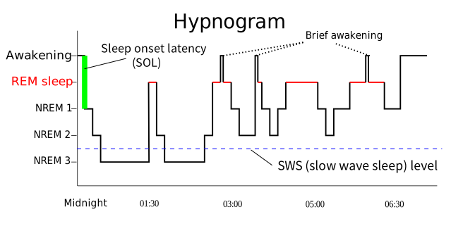
Taylor 10e (Ch 35) still teaches four NREM stages, with the same percentages and the same I–II–III–IV–III–II–REM pattern. It adds: most people go through four or five cycles a night; each lasts about 90 to 110 minutes (the deck says 90–100); and a person woken at any point returns to sleep starting at stage I. The three-stage N1–N3 scheme is the current American Academy of Sleep Medicine scoring standard, not something either course source uses. Two citation notes: the deck’s slide 8 cites “Taylor box 33.1” and slide 12 cites “Taylor Box 34-2” — numbers from older editions (the deck’s reference list is the 9th edition). In the 10th edition they are Box 35-1 (characteristics of NREM and REM) and Box 35-2 (developmental patterns of sleep).
What Gets in the Way
Jeopardy asked for “factors that interfere with any stage of the sleep cycle” and answered alcohol, caffeine, exercise, stress, medications, shift work. Every one of those six is on slide 12, which is the fuller list:
| Factor | The deck’s example or point |
|---|---|
| Developmental (age) | Sleep needs change across the lifespan (Taylor: older adults get less stage IV and shorter REM periods). |
| Psychological stress | Anxiety, fear, grief (slide 23). |
| Motivation | “Late night parties.” |
| Culture | Bedtime rituals; position; place. |
| Life events, lifestyle, habits | Shift work. |
| Physical activity and exercise | Slide 23: avoid exercise within 2 hours of bedtime. |
| Dietary habits | Alcohol, caffeine, meals at HS (bedtime); slide 23 adds heavy meals. |
| Smoking | Nicotine is a stimulant. |
| Environment | City versus country — what is usual for this person; any environment other than home, i.e. the hospital. |
| Illness | Reflux, pain, hypothyroidism. |
| Medications | Listed without examples; slide 23 adds drug dependency and withdrawal. |
Taylor on alcohol: a drink may help someone fall asleep, but larger amounts limit REM and delta sleep, and sleep fragments once the alcohol is cleared. That is why alcohol is on the “limit at bedtime” list even though patients often describe it as a sleep aid.
How much sleep, by age — Taylor 10e (Chapter 35), not the deck, which says only that needs change with development. Newborns average about 16 hours in 24; infants 12–16 (with naps); toddlers 11–14; preschoolers 10–13; school-aged children 9–12; teenagers 8–10; adults 7 or more; older adults 7–8 (up to 9). Older adults need about as much as younger adults but get it in lighter, more broken sleep, with less stage IV and shorter REM. The deck’s rule still decides an exam item: judge a patient’s sleep against their own usual pattern.
Shift Work
Shift work disrupts the circadian rhythm by making the body sleep when its clock says wake. Slide 22 lists the possible adverse effects: anxiety and depression, GI symptoms, chronic fatigue, and a set of long-term health risks the deck marks with an arrow.
An up-arrow is drawn in front of “health Risks—colorectal/breast CA; HTN, CV disease; substance abuse”: shift work raises the risk of colorectal and breast cancer, hypertension, cardiovascular disease and substance abuse.
Notice how much it overlaps slide 5’s red list for chronic sleep loss — hypertension and heart disease are on both. Shift work is chronic sleep disruption with a schedule attached.
The Joint Commission issued a sentinel alert in 2011 acknowledging the impact of fatigue on healthcare workers, with suggestions to reduce the risk:
- Assess work schedules — address extended shifts and hours.
- Review hand-off processes for safety.
- Give staff input into designing schedules that minimise fatigue.
- Educate staff on sleep hygiene and the effect of fatigue on safety.
- Address staff concerns; encourage teamwork.
- Consider fatigue when reviewing adverse events.
When Sleep Goes Wrong
The deck sorts sleep disorders into two classes. A dyssomnia is a problem with the amount, timing or quality of sleep itself — too little, too much, or broken. A parasomnia is a behaviour that happens during sleep — the sleep may be fine, but something is done in it.
| Disorder | Class | What the deck says |
|---|---|---|
| Insomnia | Dyssomnia | The most common. Difficulty falling asleep, intermittent sleep, or early wakening. Acute = a few days; chronic = a month or more. Women and older adults are more susceptible. |
| Hypersomnia | Dyssomnia | Excessive sleep, particularly during the day. |
| Narcolepsy | Dyssomnia | An uncontrollable desire to sleep that can happen at inappropriate times — risk for injury. |
| Sleep apnea | Dyssomnia | Breathing stops repeatedly in sleep — see the blue box below. |
| Restless leg syndrome | Dyssomnia | Unable to lie still because of a sensation in the legs. |
| Sleep deprivation | Dyssomnia | A decrease in amount, consistency or quality of sleep. |
| Somnambulism | Parasomnia | Sleepwalking (a stage IV event). |
| Sleep talking | Parasomnia | Also stage IV. |
| Enuresis | Parasomnia | Urinating during sleep (stage IV). |
| Bruxism | Parasomnia | Grinding teeth. |
| Nocturnal erections | Parasomnia | Listed by the deck as a parasomnia. |
| Sleep-related eating disorder | Parasomnia | Eating while asleep. |
Definition (italic on the slide): in an hour, more than 5 cessations of breathing, each lasting 10 seconds. Then, in red: “saturation drops – awakens – back to sleep – always tired?” The result is decreased arterial oxygen saturation.
Two kinds: central — CNS dysfunction in the respiratory control centre of the brain, which fails to trigger breathing during sleep; obstructive — the structures of the mouth and throat relax during sleep and occlude the upper airway.
In red: “Diagnosed with Sleep studies.”
Taylor 10e defines adult obstructive sleep apnea as five or more obstructive events per hour with symptoms (daytime sleepiness, loud snoring, observed apnea, gasping), or more than 15 per hour even without symptoms; pauses last 10–20 seconds and can reach 2 minutes. The sleep study is a polysomnogram (EEG, eye movements and muscle tone recorded overnight). Treatment is commonly CPAP. On the exam, use the deck’s wording: more than 5, lasting 10 seconds, in an hour.
Three review-sheet questions
| Teach | Because |
|---|---|
| Exercise regularly | Activity keeps alertness up. |
| Eat small meals high in protein | Protein wakens the brain (slide 24, red) — and a small meal avoids the drowsiness of a heavy one. |
| Avoid what increases sleepiness — alcohol, sitting too long, warm temperatures | Each one invites an attack. |
| Avoid what could cause injury — driving while drowsy, heights such as a ladder | Falling asleep there is the injury risk. |
| Take naps when drowsy or when events are likely | A planned nap is safer than an unplanned attack. |
| Take prescribed stimulants | The medical treatment. |
Assessing Sleep
The sleep assessment is mostly subjective — the patient’s own account — with a few things you or a bed partner can observe. The deck gives it in four pieces.
| Piece | What to cover |
|---|---|
| Nursing interview — assess history (slide 17) | The patient’s usual sleep–wake pattern and recent changes; the effect on everyday functioning and usual sleep requirement; use of sleep aids; sleep disturbances and contributing factors. Have them rate their sleep 0–10, best to worst. |
| If there is a disturbance (slide 18) | Its nature and cause; accompanying signs and symptoms; when it began and its effect on everyday living; severity; treatment so far; how the patient is coping. |
| Characteristics to observe (slide 19) | Restlessness, sleep postures, sleep activities, snoring, leg jerking. |
| Sleep diary (slide 20) | Time the patient retires; time they try to fall asleep; approximate time they fall asleep; any awakenings and return to sleep; time of waking in the morning; stressors; food, drink, medications; physical and mental activities 2–3 hours before bed; worry. |
Helping Patients Sleep
Slide 23 frames the nursing problem — sleep disturbance, showing up as difficulty remaining asleep, premature awakening, excessive daytime sleeping or an altered sleep–wake pattern — and lists the usual causes: emotional stress, exhaustion, a changed bedtime ritual or environment, circadian disruption from travel or work, exercise within 2 hours of bed, caffeine and heavy meals, drug dependency and withdrawal, and physical illness.
Treating a dyssomnia (slide 24)
| Kind | What the deck lists |
|---|---|
| Nonpharmacologic — first | Stimulus control; sleep restriction; sleep hygiene; cognitive therapy; multicomponent therapy; relaxation therapy. |
| Pharmacologic — last resort | Sedatives and hypnotics: “Use as last resort.” |
Standard nursing content, in brief: stimulus control means using the bed only for sleep and getting up if not asleep after a while; sleep restriction limits time in bed to the time actually spent asleep, then lengthens it. Taylor names cognitive behavioural therapy as the first-line treatment for chronic insomnia.
| Intervention (slide 25) | At the bedside |
|---|---|
| Prepare a quiet, restful environment | Limit noise, dim the lights, lower the room temperature (Jeopardy’s three). |
| Promote bedtime rituals | Keep the patient’s own routine — reading, washing, a set position. |
| Offer appropriate bedtime snacks and beverages | A light carbohydrate snack — see the box below. |
| Promote relaxation and comfort | Pain relief, positioning, a clean dry bed. |
| Respect normal sleep–wake patterns | Work with this patient’s usual bedtime, not the unit’s. |
| Schedule nursing care to avoid disturbances — “allow time for REM sleep” | Group care so the patient gets unbroken blocks of at least one 90–100-minute cycle; REM comes at the end of each cycle. |
| Use medications to produce sleep | As ordered — and as the last resort (slide 24). |
| Teach about rest and sleep | Sleep hygiene; the factors on slide 12. |
| Limit alcohol, caffeine, nicotine at HS (bedtime) | Caffeine and nicotine are stimulants; alcohol fragments sleep. |
| Limit fluids 2–4 hours before HS | Fewer trips to the bathroom — fewer broken cycles. |
| Source | What it says |
|---|---|
| Deck, slide 24 (red) | “Protein wakens the brain … carbohydrates stimulate melatonin.” |
| Review sheet | “Which is the best to prevent sleep disturbance? Avoid caffeine, spicy foods, and protein? Bland food?” |
| Jeopardy | Promotes sleep: “a snack of cereal and milk.” Avoid before bed: “chocolate, cheese, or french fries.” |
So the best bedtime snack is light, bland and carbohydrate-based — cereal and milk, toast, crackers. Each of Jeopardy’s “avoid” foods fails for its own reason: chocolate has caffeine, cheese is protein, French fries are heavy and fatty.
Taylor disagrees on protein: it recommends a small protein-and-carbohydrate snack (it even suggests “cheese and crackers” or peanut butter on toast) because protein supplies L-tryptophan. The course sources all say avoid protein and cheese at bedtime, so answer with the course. Taylor also advises avoiding caffeine for 4–5 hours before bed and avoiding large, heavy or spicy meals — which agrees with the course.
The Week’s Terms
Both preparation sheets end with a “Define Terms” list. Here they are together, each in one plain line, using the decks’ own definitions wherever the decks give one.
| Term | In one line |
|---|---|
| Apex of the heart | The bottom — the tip of the left ventricle, at the 5th left ICS, midclavicular line. (Lungs are named the other way round.) |
| Base of the heart | The top, where the great vessels attach; the aortic and pulmonic areas, where S2 is louder. |
| Dysrhythmia | “Abnormality of rhythm” (deck) — an electrical malfunction. Jeopardy says arrhythmia; same thing here. |
| Systole | “Myocardial contraction” — ejection of blood. Starts with S1. |
| Diastole | “Filling phase” — the heart relaxes and fills; the coronary arteries fill now too. Starts with S2. |
| Murmur | A blowing or swooshing sound when blood volume is increased or flow is impeded or altered. Systolic just after S1, diastolic just after S2. |
| Preload | “Degree of stretch on the ventricle” — how full it is before it pumps. |
| Afterload | “Pressure the ventricle has to pump against.” |
| Palpitation | The client’s awareness of irregular, skipped or pounding beats — subjective. |
| Jugular vein distention (JVD) | Bulging neck veins — venous congestion backing up from the right side of the heart. |
| Point of maximal impulse (PMI) | Where the heartbeat is best felt on the chest wall — usually the 5th ICS, midclavicular line. The deck drew an arrow to this one. |
| Edema | Swelling from fluid in the tissues; graded +1 to +4 by pit depth. A cue for venous trouble or heart failure. |
| Atherosclerosis | “Narrowing from plaque” (PV deck) — the most common cause of PAD and of coronary disease. |
| Arteriosclerosis | “Thickening/hardening of arteries” (PV deck) — calcification with age; raises systolic BP. |
| Thrombus | A clot that forms and stays in a vessel — often where the endothelial lining is injured. |
| Embolism | A clot (or other mass) that has broken loose and travelled, then blocks a vessel downstream — a DVT that reaches the lung is a pulmonary embolism. |
| Deep vein thrombosis (DVT) | A thrombus in a deep vein — more serious than a superficial one because of the risk of pulmonary embolism. Warm, swollen, tender limb. |
| Varicosities | “Distended, protruding veins that appear darkened and tortuous” (tidbits) — weakened walls and valves. |
| Angina | Chest pain or pressure from too little oxygen to the heart muscle — chest, arm, jaw; stable or unstable. |
| Bruit | A swishing sound over an artery, heard with the bell — narrowing or disrupted flow. |
Thrombus and embolism are listed but not defined in the decks; those two lines are standard definitions, consistent with the tidbits handout’s use of the words.
The marked items, one more time
| Item — and what marks it | Answer |
|---|---|
| The heart on a skeletal figure — eight questions | |
| APETM, with rib addresses — “You will be tested on this” (APETM lab activity, heart lab prep, blueprint) | Aortic 2nd ICS RSB, ribs 2–3 · Pulmonic 2nd ICS LSB, ribs 2–3 · Erb’s point 3rd ICS LSB, ribs 3–4 · Tricuspid 4th ICS LLSB, ribs 4–5 · Mitral 5th ICS left MCL, ribs 5–6. Sternal angle = rib 2; a space is named for the rib above it. Give letter, full name, space and border |
| Where S1 and S2 are loudest — “where S1S2 heard loudest and why (will be tested)” (heart lab prep, APETM lab activity, blueprint) | Base (A, P) S2 louder · Erb’s S1 = S2 · apex (T, M) S1 louder, loudest at the mitral area. Why: the sites are near, not over, the valves; sound radiates with the flow |
| PMI and the apical pulse — cardiac deck, drawn arrow (slide 16); blueprint, “apical pulse and location” | 5th ICS, midclavicular line = apex = apical pulse = mitral area. Rate and rhythm first, at the apex or Erb’s point (the alternative apical site): regular, 30 s × 2; irregular, a full minute |
| Valve location vs listening post — the “and why” (heart lab prep; deck notes on the APETM slide) | Valves sit together behind the sternum; APETM sites are listening posts, downstream. Two cross the midline: aortic valve left of centre, heard 2nd right ICS; tricuspid right of centre, heard lower left sternal border. “Heard” or a figure → the post |
| Diastole · systole — cardiac deck, block arrows (slide 20) | Diastole → filling. Systole → ejection of blood. S1 = mitral + tricuspid shut, start of systole; S2 = aortic + pulmonic shut, start of diastole |
| The path — “Memorize/understand the blood flow” (heart lab prep); “Important to understand blood flow and internal structures” (blueprint) | “Venae cavae, right atrium, tricuspid, right ventricle, pulmonic, pulmonary arteries, lungs. Pulmonary veins, left atrium, mitral, left ventricle, aortic, aorta, body.” It is how every backup question is answered |
| Cardiac — the blueprint’s lines and the deck’s only red text | |
| Chest pain — cardiac deck, red text (slide 14, repeated on 23) | “Consider all chest pain to be cardiac until cardiac issue is ruled out.” Gather data while treating with O2 and nitroglycerin |
| Chest pain, first question · what it is called — blueprint; prior-year review | Have the client describe the pain — “the most important and first additional information.” Pain in the chest, arm or jaw = angina, stable or unstable (Jeopardy); “cells deprived of O2” |
| Pericardial or pleural rub — blueprint; Jeopardy, twice | Ask the client to hold their breath. Pleural rub stops; pericardial rub keeps going, because the heart keeps beating |
| “Causes for extra sounds like S3 and S4” — blueprint | S3, ventricular gallop, “Ken-tuc-ky”, after S2: an overfull ventricle — heart failure, fluid overload. S4, atrial gallop, “Ten-nes-see”, before S1: a stiff ventricle — CAD, HTN, MI, aortic or pulmonic stenosis |
| Blood not moving forward, left and right — blueprint | Left → lungs: crackles at the bases, dyspnea, orthopnea, cough. Right → venae cavae → body: JVD, dependent edema, weight gain, enlarged liver. Tricuspid fails → cavae; high pulmonary artery pressure → right ventricle |
| Why nocturia — blueprint | Fluid pools in the legs by day; lying flat returns it; more blood reaches the kidneys, so more urine overnight. With edema → heart failure |
| Decreased cardiac output — blueprint | Decreased tissue perfusion: falling BP, LOC changes, less urine with rising BUN and creatinine, cool pale skin, slow capillary refill, fatigue. The priority vital sign is BP |
| Health promotion / risk reduction — blueprint | Modifiable: smoking, BP, cholesterol, weight, glucose, inactivity and diet, stress. Teach from “what the patient is willing to work on” (Jeopardy) |
| Both echocardiograms — blueprint | Same ultrasound, different doorway. Transthoracic: gel and transducer on the chest, non-invasive. Transesophageal: probe down the oesophagus, behind the heart |
| Stress test — blueprint, “Meds that slow heart rate” | Hold heart-rate-slowing meds (beta blockers) as the provider directs · no tobacco or caffeine · comfortable clothing |
| Lipid profile — blueprint, “good and bad cholesterol”; deck notes, HDL “GREATER” | LDL is the bad one, the most dangerous. Total <200 · LDL <100 · HDL >40 · triglycerides <150. HDL is the only one you want high |
| The carotid — blueprint, “cause of abnormal findings” | Palpate one side at a time. Listen with the bell. A bruit = narrowing or disruption of flow: plaque, or an enlarged thyroid. A carotid thrill → listen for a bruit |
| Peripheral vascular — said in the lecture, set in red, named by the blueprint | |
| Pulse grade — lecture: “a test question on a previous test” | Normal is 2+, documented 2+/4. 0 absent · 1+ weak · 3+ full · 4+ bounding |
| Capillary refill — said in the lecture | “Two to three seconds” — under 3. Longer is delayed |
| Why each pulse site — lecture: “you should know this”; blueprint, “names of pulses” | Radial routine · temporal if radial not accessible · carotid adult arrest · apical infants and children, and a discrepancy with the radial · brachial BP and infant arrest · femoral arrest and shock · popliteal lower leg · posterior tibial and dorsalis pedis the foot |
| SCDs — PV deck slide 10 in capitals; notes underlined; said in the lecture | “How can we enhance venous return in hospitalized patients?” SCDs simulate the calf-muscle pump for bed-bound and post-surgical patients |
| Arterial occlusion — PV deck, red text (slide 13) | Pain, numbness and tingling, coolness or colour change = urgent, possible total arterial occlusion |
| The P’s — tidbits handout bold, “Know the 6 Ps”; blueprint, “arterial insufficiency and occlusion” | Pain, pallor, pulseless, paresthesia, polar, paralysis, perfusion (capillary refill). Learn seven; six drops perfusion. Chronic insufficiency: claudication, hair loss, thick nails, cool pale skin, pallor raised, rubor dependent, weak or absent pulses, painful toe ulcers |
| One limb cooler, or one warm and swollen — blueprint, “Normal/Abnormal findings”; PV deck slide 21, read aloud in the lecture | One cooler = arterial occlusion. Warm, edematous, tender = DVT (30–40%). Both emergent. Compare limb to limb, in a warm room |
| Preventing venous emboli — blueprint; review sheet | Ambulation · SCDs · anti-embolism stockings · elevate the legs while sitting · no crossed legs, no constrictive clothing · avoid long sitting or standing · ROM · check DP and PT pulses · stop smoking · anticoagulants as ordered |
| Teaching for venous disease — blueprint | Elevate (above the heart in bed; 10–20 minutes every few hours) · stockings on before getting out of bed · no long sitting or standing, crossed legs or tight clothing |
| Arterial risk and prevention — blueprint | Smoking, “one of the most devastating” · diabetes, four times the risk of PAD · most modifiable: smoking, high-fat diet, limited activity. Non-modifiable: genetics, age, gender, ethnicity |
| Sleep — six questions, “PP slides” | |
| Why sleep matters — sleep deck, red text (slides 4, 5) | Rest and sleep are critical for physical, mental and emotional wellbeing. Chronic sleep loss → obesity, depression, hypertension, diabetes, heart attack, stroke |
| REM and NREM — sleep deck, drawn arrows (slide 8); blueprint | NREM ↓ temperature, HR, RR, BP, metabolic rate (parasympathetic) · REM ↑ the same five, varying, with ↓ muscle tone and reflexes. NREM 75–80%, four stages; REM 20–25%, vivid dreams, harder to arouse than NREM. Stage IV: delta, deepest — sleepwalking, sleep talking, enuresis |
| Normal sleep cycle — blueprint | I → II → III → IV → III → II → REM, 90–100 minutes; first REM about 90 minutes in; the next cycle re-enters at stage II |
| Chemicals present — blueprint; sleep deck, red text (slide 24) | Melatonin regulates the sleep–wake cycle; the hypothalamus is the control centre. “Protein wakens the brain … carbohydrates stimulate melatonin” → bedtime snack cereal and milk |
| Sleep apnea — sleep deck, red text (slide 15) | Saturation drops – awakens – back to sleep – always tired. Diagnosed with sleep studies. (The definition, in italics: more than 5 cessations of breathing, each lasting 10 seconds, in an hour) |
| Shift work — sleep deck, drawn up-arrow and red text (slide 22) | Shift work ↑ colorectal and breast cancer, hypertension, CV disease, substance abuse. Joint Commission sentinel alert on fatigue in healthcare workers |
| Nursing sleep interventions — blueprint | Quiet restful room · bedtime rituals · carbohydrate snack · relaxation and comfort · respect the usual pattern · schedule care to allow time for REM · medication as a last resort · teach · limit alcohol, caffeine, nicotine · limit fluids 2–4 hours before bed |
The night before: the first five rows of the box are the eight figure questions, and the sixth is the chain every backup question rests on — say every site with its space, its rib address and its louder sound, out loud, top to bottom. The last seven rows are the six sleep questions. Everything between is the thirty-two multiple choice.
Other things worth knowing
| Item | Answer |
|---|---|
| The four valves, in order | Tricuspid, pulmonic, mitral, aortic — each the exit door of the chamber before it |
| Two pumps | Same volume every beat; the left is stronger, not busier |
| Where the colour changes | Red in the lungs, blue in the body — never inside the heart |
| Thickest wall | Left ventricle — the tell on an unlabelled drawing |
| Base · apex | Top · bottom — the opposite of the lungs |
| Other names | Mitral = bicuspid = left AV · tricuspid = right AV · pulmonic = pulmonary |
| Apex in a word bank | A landmark, not a stop |
| The drill | Eleven with the bank → bank hidden → the nine → Trace it |
| Pulmonary artery or arteries | The blank cares which vessel, not right, left or plural |
| Item | Answer |
|---|---|
| Systole · diastole, deck words | “Myocardial contraction” · “filling phase”; relaxing = diastole (Jeopardy) |
| Which sound is S1 | The one after the long pause |
| Coronary arteries | Fill in diastole — a fast heart shortens it |
| S3 · S4, when normal | S3: children, late pregnancy, high output; rarely normal in older adults; heard best left lateral. S4: athletes, elderly; “usually abnormal” |
| S1, S2 and S3 | Heart failure |
| Murmurs | Blowing. Systolic after S1, diastolic after S2; graded 1–6; diastolic or above 3/6 never innocent |
| Murmur · bruit · thrill · rub | Heard over the heart = murmur · over an artery, bell = bruit · felt = thrill (then listen there) · scratchy, through a held breath = pericardial rub |
| Other extra sounds | Split S2: normal, pulmonic area, inspiration · ejection click: great arteries distending · opening snap: mitral stenosis |
| Pulse deficit | Apical − radial, irregular pulse, a full minute |
| Draping | Displace breast tissue laterally; expose only the part assessed |
| Item | Answer |
|---|---|
| The one rule | Backs up one stop back, goes short one stop forward; mitral → lungs, tricuspid → cavae |
| Orthopnea | Breathless lying flat: “How many pillows do you use for sleep?” |
| Right-sided edema | Dependent, usually both sides: feet and ankles, sacrum in bed |
| Weight gain | About a kilogram a litre — shows before the ankles. Daily weights |
| Commonest cause of right-sided failure | Left-sided failure |
| Cardiac output | SV × HR; preload = stretch, afterload = pressure pumped against. SV falls → HR rises |
| The deck’s three problems | Impaired cardiac output, impaired tissue perfusion (forward) · excess fluid volume (backward) |
| Item | Answer |
|---|---|
| The bolded history list (slide 10) | Angina (chest, arm, jaw) · palpitations · fatigue · difficulty breathing · orthopnea · dizziness · nocturia · edema · heartburn/indigestion · diaphoresis |
| Sort the list | Muscle short of oxygen? Rhythm wrong? Pump failing, forward or backward? |
| The two “twice”s | High LDL and diabetes each make cardiovascular disease twice as likely (deck notes) |
| Medications | What, why, taken as prescribed — and OTC and supplements. HB’s weekly antacids were the clue |
| Pillows · up at night | Orthopnea and nocturia — heart-failure screens asked as sleep questions |
| Slide 14’s differential | MI, angina, GI, pulmonary/pleural, musculoskeletal, aortic dissection, pericardial — assume cardiac while you sort them |
| The first minutes | Assume cardiac → describe the pain → BP → O2 and nitroglycerin while gathering data → notify → reassess pain and BP → document last |
| Women (slide 24) | “Present differently than men”; underrepresented in research. No crushing pain does not rule out the heart |
| Modifiable or not | HTN and glucose are modifiable. Genetics, family history, age, gender, ethnicity are not |
| Slide 9 — reduce CHD deaths | Smoking cessation · warning signs, call 911 · AEDs · HTN control · lower cholesterol · office visits for co-morbidities · vigorous activity |
| What motivates | The patient’s own goal — not a list of consequences |
| Equipment · preparation | Stethoscope, watch with a second hand · explain, privacy, gown, expose only what is assessed, blanket |
| Inspect · palpate · percuss | Lesions, masses, deformity, JVD, PMI · PMI, carotid, masses · percussion “not commonly performed” |
| PMI not palpable in an adult | Not abnormal by itself; pulsations anywhere else are |
| Listening positions | Sitting, supine, left lateral recumbent (lab manual); bell and diaphragm at every site; S3 best heard left lateral. Palpate a thrill → listen for a murmur |
| Carotid | One at a time, “to ensure adequate cerebral blood flow”. Used in adult arrest; femoral if it cannot be felt |
| Carotid or jugular (lab manual) | Jugular: undulating, easily obliterated by light pressure, changes with position and breathing. Carotid: one positive wave. JVP at 45° under 3 cm; negative abdominojugular (hepatojugular) reflux is normal |
| Which test for which question | Muscle damage now → troponin, CK-MB · failing → BNP · long-term risk → lipids, homocysteine, CRP · kidneys → BUN, creatinine |
| ECG · Holter · catheterization | Electrical conduction · ECG over a prolonged period · diagnoses coronary blockages |
| SA node (slide 7) | Fires the impulse that starts each beat — “the conduction system tells the mechanical system when to beat” |
| Asystole (Jeopardy) | “Flatline” — no cardiac output. An arrhythmia is an electrical malfunction of the heart |
| After a TEE | Nothing by mouth until the gag reflex returns (standard nursing content) |
| Clinical judgement (slide 25) | Identify cues → analyse, hypothesise → prioritise → generate solutions → take action → evaluate |
| Medications | “Learn by classification” — diuretics, antiplatelets, anticoagulants, beta and calcium channel blockers, ACE inhibitors, anti-anginals, thrombolytics |
| HB, the cardiac case | 72, anterior MI, 10/10 “pressure” he called indigestion. Crackles, RR 28, HR 115 irregular, BP 170/105, S3, JVD. Priority: decreased output / cardiac perfusion, then excess fluid volume |
| Item | Answer |
|---|---|
| Hardening · plaque | Arteriosclerosis · atherosclerosis — the one pair the lecture separated |
| Thrombus · embolus | Stays · travels. A DVT that reaches the lungs is a PE |
| Arteries · veins | Thick, elastic, high pressure, driven by the heart · thin, low pressure, driven by muscle, a pressure gradient and valves |
| Superior · inferior vena cava | Where a PICC tip sits · where a filter goes |
| Venous return | Pressure gradient · one-way valves · calf muscle pump |
| Lifespan | Pregnancy: blood volume up (the deck: “doubles”), leg edema expected · older adults: stiff arteries → systolic rises · prevention begins in childhood |
| Cramping when walking | Intermittent claudication; hair loss, pale cool skin = poor arterial supply; edema = venous |
| Why the room is warm | “Cold temps make vasoconstriction” — false positives. Compare one side with the other |
| Hand hygiene (Jeopardy) | Every time, on entering and on leaving the patient’s room |
| Pitting edema | +1 to +4 = 2, 4, 6, 8 mm — grade × 2. No “normal” grade |
| Temperature | Backs of the hands, both limbs at once |
| Homans’ sign | No longer recommended — unreliable, and may dislodge a clot |
| Posterior tibial | Behind and below the medial malleolus |
| One foot pulse, four names | Pedal = dorsal pedal = dorsal = dorsalis pedis, top of the foot |
| Two +4 scales | Pulse 2+ = normal; edema 2+ = abnormal; pulse 4+ = bounding, abnormal |
| Lab-manual pulse scale | Lists 0, 1, 3, 4 and skips 2 — answer with the deck: 2+/4 is normal. Allen test if indicated: arterial flow to the hand |
| Doppler | Gel, transducer slightly angled over the artery, volume up. Rhythmic whoosh = blood getting through |
| Carotid and jugular, listening | Bell, breath held. Adult: no bruit. A venous hum disappears when you press on the jugular; may be normal in children |
| BP (lab-manual table) | Both arms; supine, sitting, standing · palpate, then inflate 30 mm Hg above · pulse pressure ≈ ⅓ of systolic · sounds to “0” → record Korotkoff 1, 4, 5 · its orthostatic 10–15 and <140/90 are older cut-offs |
| Labs | Arterial: lipids, HbA1c, CRP, homocysteine (risk) · venous: D-dimer, venous Doppler/duplex (the clot) |
| Slide 31 hypotheses | Altered tissue perfusion, arterial · risk for peripheral neurovascular dysfunction · activity intolerance |
| Slide 31 goals | Pulses strong and symmetrical · refill <3 seconds · edema down · senses sharp and dull · teach-back |
| Keep limbs warm | Socks, blankets, a warm room — never a heating pad |
| Cold, pale, pulseless foot | Assess (Doppler if needed, compare sides) → report at once → document. Do not raise, rub or heat it |
| Item | Answer |
|---|---|
| PAD · PVD (tidbits handout) | Arterial side, starved · venous side, flooded |
| Arterial leg | Pale, cold, hairless, thick nails, weak or absent pulses, little edema, painful ulcer on the toes or top of the foot |
| Venous leg | Swollen, warm, brown staining from ankle to calf, aches after standing, ulcer around the ankle |
| Rest pain | Later than claudication; wakes at night; eased by dangling the leg |
| PAD exercise | Walk to the point of claudication, rest, walk a little farther — collateral circulation |
| PAD warmth | Insulated socks and shoes; avoid cold; no heating pad — possible neuropathy |
| Acute occlusion | Sudden, one limb colder and paler, pulseless → notify now; embolectomy or thrombolytic |
| After embolectomy | Compare the P’s with the pre-op baseline. Worsening pain → compartment syndrome or re-occlusion |
| Raynaud’s · Buerger’s | Vasospasm with cold or stress, white → blue → red · inflammatory occlusion, constant, stop smoking first |
| Clot words | Thrombophlebitis = clot + inflammation · phlebothrombosis = clot, no inflammation · phlebitis = IV-related inflammation |
| DVT care | Bed rest as ordered · no massage, no pillow under the knee · elevate · measure the circumference |
| Heparin · Coumadin | Stops growth, PTT · longer-term, PT/INR. A thrombolytic dissolves the clot |
| IVC filter | Traps emboli; does not dissolve the clot. New back pain + changing vitals → retroperitoneal bleed |
| Best single VTE preventer | The one that moves the calf: ambulation, or SCDs if they cannot walk |
| Unna boot | Venous treatment; too tight → arterial occlusion |
| Varicose veins | Dull ache, fullness after standing, ankle edema — stockings, elevate, no crossed legs |
| Valve competence | Trendelenburg test or manual compression test if varicosities are present (lab manual) — not the Trendelenburg position |
| Neuropathy · lymphedema · AAA | Numb feet, diabetes · lymph cannot drain · ballooned abdominal aorta, a bruit site |
| JH, the PV case | 55, diabetes, HTN, CAD; claudication, absent left pedal pulses, toe ulcers, no edema — arterial, chronic. First problem: altered tissue perfusion, arterial |
| Item | Answer |
|---|---|
| Rest · sleep | Less activity, refreshed · altered consciousness, restores mental and physical function |
| Short-term loss | Poor concentration and judgement, irritability, accidents; ↓ leptin → ↑ appetite |
| The brain’s parts | RAS and bulbar synchronizing region (brainstem) cycle sleep · hypothalamus controls it; injured → abnormally long sleep |
| Circadian rhythm · sleep cycle | 24 hours · 90–100 minutes. Light and dark influence the clock, “not primary control” |
| Stage shares | I ≈ 5% · II ≈ 50% · III and IV 10% each · REM 20–25% |
| REM rebound | Lose REM and it increases on later nights |
| Shape of the night | Deep sleep early; REM longer towards morning |
| What interferes (Jeopardy) | Alcohol, caffeine, exercise, stress, medications, shift work |
| Exercise | Not within 2 hours of bedtime |
| Dyssomnia · parasomnia | The sleep itself is wrong (insomnia, narcolepsy, apnea, RLS) · something done in sleep (sleepwalking, bruxism, enuresis) |
| Apnea types | Central: brain fails to trigger · obstructive: airway relaxes shut, snoring |
| Sleepwalker who fell | Alarm the bed |
| Narcolepsy | Anticipate falling asleep at odd times → risk for injury. Small high-protein meals, planned naps, no driving drowsy, stimulants |
| Assessment | Usual pattern first, then the change; rate sleep 0–10; sleep diary separates in bed from asleep |
| Treating a dyssomnia | Non-drug first; sedatives and hypnotics “as last resort” |
| Jeopardy’s environment | Limit noise, dim lights, lower the room temperature |
| Bedtime snack | Light carbohydrate — cereal and milk. Not chocolate (caffeine), cheese (protein), French fries (heavy) |
Traps, one line each
| Trap | Correct answer |
|---|---|
| “Arteries are oxygenated” | Arteries leave the heart; the pulmonary arteries are blue |
| Labelling by the side of the page | Patient’s right is on your left |
| S1 as the filling sound | S1 starts systole; filling is the silence after S2 |
| S4 as “ventricular gallop” (slide 21) | S4 is the atrial gallop |
| S4 “normal in children” (slide 3) | It is S3 that is normal in children |
| Cough to sort a rub | Cough is for crackles and rhonchi. Rub → hold the breath |
| Aortic valve heard on the left | 2nd ICS, right sternal border |
| “Located” vs “heard” | Located = behind the sternum; heard = the listening post. A figure wants the post |
| Counting ribs from the top of a drawing | Start at the sternal angle = rib 2 |
| “Hear a thrill” | A thrill is felt; a swish over the carotid is a bruit, not a murmur |
| Tricuspid area per the lab manual (“S1 > or = S2”) | Use the deck: S1 louder at tricuspid; S1 = S2 only at Erb’s |
| PMI at the “4th–5th ICS” (slide 16 notes) | Answer 5th |
| High pulmonary artery pressure | Overworks the right ventricle |
| Crackles in a cardiac stem | Left-sided failure before pneumonia |
| Edema always means the heart | One warm, swollen, tender leg = DVT; both legs plus JVD = heart |
| Decreased output means BP is already low | Watch BP because it will show the fall; nitroglycerin can drop it too |
| Shiny, hairless legs after an MI | Months of arterial insufficiency, not the MI |
| The pain score as the first question | “Describe what it feels like” comes first |
| Better after an antacid, so GI | Relief is not a diagnosis. Rule out the heart first |
| No crushing pain, so not cardiac | Women and older adults present differently |
| Hypertension and diabetes are non-modifiable | Modifiable — they can be controlled |
| Diabetes: twice or four times? | Twice the cardiovascular disease (cardiac deck) · four times the lower-extremity PAD (PV deck) |
| Warn of consequences to motivate | Start from what the patient is willing to work on |
| PMI not felt → notify | “May or may not be palpable” in adults |
| Both carotids together | Never — one at a time |
| Normal total cholesterol clears the patient | Read LDL; and an HDL under 40 is bad |
| “Stop all your heart pills” | Hold only the rate-slowing ones, as the provider directs |
| “Denies shortness of breath” at RR 28 | Document both; act on the measurement |
| Capillary refill “less than 2 seconds” (Jeopardy) | The instructor said two to three seconds; hers wins |
| Six P’s or seven | Learn seven; six is seven without perfusion |
| “Claudication — pain at rest” (PV slide 30) | Claudication is pain with walking, eased by rest; rest pain is a later sign |
| Test Homans’ sign, massage the calf | Neither — may dislodge a clot |
| Pulse 2+ and edema 2+ mean the same | Pulse 2+ is normal; any pitting is abnormal |
| Doppler “whoosh” is a bruit | The Doppler whoosh is the normal arterial signal |
| A carotid bruit proves plaque | Plaque or an enlarged thyroid — it names turbulence, not the cause |
| Pale, cool feet in a cold room | Warm the room, recheck, compare sides |
| Heating pad for cold PAD feet | Never — socks and shoes |
| Raise every bad leg | Venous: up, above the heart in bed. Arterial: a little, in a chair, never above the heart; dangle in severe PAD |
| Pillow under a DVT knee | Kinks the vein — elevate the whole leg |
| The IVC filter treats the DVT | It traps emboli; it does not dissolve the clot |
| “Limit fluid?” for vascular disease | Not a course intervention; fluid restriction belongs to heart failure |
| Checking pulses prevents VTE | It is assessment; moving the calf is prevention |
| Worse pain after embolectomy is surgical | Compartment syndrome or re-occlusion — report |
| Teach JH to stop smoking | The case never says he smokes — assess, do not assume |
| Fast, irregular pulse in a sleeping patient | Recognise REM first |
| Sleepwalking in REM | Stage IV — muscles are limp in REM |
| “Stage I and II — 5% & 50%” as a range | Two numbers: stage II is about half the night |
| Hardest to wake | REM vs NREM → REM; deepest NREM stage → stage IV |
| Three NREM stages, N3 | Answer with the deck’s four; N3 = III and IV |
| Circadian rhythm vs sleep cycle | 24 hours vs 90–100 minutes |
| Exercise ruins sleep | Only within 2 hours of bed |
| Quiet room = good environment | What is usual for this patient |
| Narcolepsy as a parasomnia | Dyssomnia — the problem is when and how much |
| Cheese and crackers at bedtime | Cheese is protein — cereal and milk |
| Sleeping pill first | Non-drug measures first; medication last |
| “Allow time for REM” = do not wake them | Cluster the care; ordered checks still happen |
| “Eight hours in bed” | Time in bed is not time asleep |
Read the recap
Recap: The Path, Drawn
Everything about the heart on this exam rests on one chain, and the blueprint says so: you need to understand blood flow and internal structures to answer these questions. Say it as two sentences. Venae cavae, right atrium, tricuspid, right ventricle, pulmonic, pulmonary arteries, lungs. Pulmonary veins, left atrium, mitral, left ventricle, aortic, aorta, body. The valves in order are tricuspid, pulmonic, mitral, aortic: Try Pulling My Aorta. Remember three things. An artery leaves the heart, so the pulmonary arteries carry blue blood. Right and left are the patient’s, so the right heart is on your left. And Apex is a landmark, not a stop. The left ventricle has the thickest wall, and blood changes colour only in the lungs and the body, never inside the heart.
Recap: Drill It
Part 2 is practice. Do the eleven blanks with the bank showing, then hide the bank and do it again. Then do the nine, which jumps from each ventricle straight to its artery. The figure questions and every backup question on Exam 4 lean on that chain.
Recap: Valves, Sounds and the Cardiac Cycle
A heart sound is a pair of valves shutting. S1, the lub, is the mitral and tricuspid closing as systole starts; S2, the dub, is the aortic and pulmonic closing as diastole starts. Diastole means filling, systole means ejection.
Eight questions on Exam 4 are a chest figure, so learn the five listening posts, top to bottom: aortic and pulmonic in the second space either side of the sternum, Erb’s point in the third, tricuspid in the fourth at the lower left sternal border, and mitral in the fifth at the midclavicular line, which is also the apex, the apical pulse and the PMI. On a drawing, start at the sternal angle: it is the second rib, and each space is named for the rib above it. S2 is louder at the base, S1 at the apex, and they are equal at Erb’s point, because the posts sit near the valves, not over them, and sound follows the flow. So the aortic valve, left of centre, is heard on the right. Take the apical rate at the apex or Erb’s point: regular, thirty seconds times two; irregular, a full minute.
S3 comes after S2, like Kentucky, and means heart failure; S4 comes before S1, like Tennessee, and means a stiff ventricle. A swoosh over the heart is a murmur, over an artery a bruit, and a thrill is felt, never heard. For a rub, ask the client to hold their breath.
Recap: When the Flow Backs Up
Work the symptoms out from the path: a failure backs blood up into the stop before it, and leaves the stop after it short. Left goes to the lungs: crackles at the bases, shortness of breath, and orthopnea, found by asking about pillows. Right goes to the rest of the body: distended neck veins, swelling in both legs, weight gain and an enlarged liver. A failing tricuspid backs up into the venae cavae, and high pulmonary artery pressure overworks the right ventricle. The commonest cause of right-sided failure is left-sided failure. Nocturia happens because lying flat returns the leg fluid to the circulation, and the kidneys clear it overnight. When output falls, every organ is short of blood, and blood pressure is the vital sign to watch, even while it still reads high.
Recap: The Cardiac Assessment
The history finds most of what the exam later confirms. The deck’s bolded list of symptoms is angina in the chest, arm or jaw, palpitations, fatigue, difficulty breathing, orthopnea, dizziness, nocturia, edema, heartburn or indigestion, and sweating. Sort them by three questions: is the muscle short of oxygen, is the rhythm wrong, or is the pump failing? Two lifestyle questions are heart-failure screens in disguise: how many pillows do you use for sleep, which finds orthopnea, and do you get up at night to urinate, which finds nocturia. Ask about over-the-counter medicines too; the patient in the cardiac case, HB, had been taking antacids every week.
Chest pain. The deck’s only red line says: consider all chest pain to be cardiac until a cardiac issue is ruled out. You gather data while you treat with oxygen and nitroglycerin. The first thing to ask is for the client to describe the pain; the review sheet calls that the most important and first information you want. Pain in the chest, arm or jaw is called angina, and it can be stable or unstable. Relief after an antacid does not rule out the heart, and women often present differently from men, so a missing classic sign rules nothing out.
Risk reduction. Smoking, blood pressure, cholesterol, weight, blood sugar, inactivity, diet and stress are the things a patient can change. High blood pressure and diabetes count as modifiable, because they can be controlled, even though they sound permanent. Genetics, family history, age, gender and ethnicity cannot change. When you teach, start with what the patient is willing to work on, not with a list of consequences.
The physical exam is inspection, palpation and auscultation; percussion is advanced and rarely done. A point of maximal impulse you cannot feel in an adult is not abnormal by itself. Palpate the carotids one at a time. For the lipid profile, LDL is the bad cholesterol and should be under one hundred; HDL should be greater than forty, the only one you want high; total cholesterol under two hundred; triglycerides under one hundred and fifty. Troponin tells you the heart muscle is being damaged now, and B-type natriuretic peptide tells you the heart is failing.
A transthoracic echocardiogram puts gel and a transducer on the chest; a transesophageal echocardiogram passes the probe down the oesophagus, behind the heart, so nothing goes by mouth afterwards until the gag reflex returns. Before a stress test, hold the medicines that slow the heart rate, such as beta blockers, as the provider directs, and avoid tobacco and caffeine. HB, seventy-two, with a ten out of ten pressure in his chest that he called indigestion, crackles, a pulse of one hundred and fifteen, an S3 and distended neck veins, was having an anterior heart attack; his first problem was decreased cardiac output and cardiac tissue perfusion.
Recap: Peripheral Vascular Assessment
The heart is the pump; the peripheral vessels are the pipes. Arteriosclerosis is hardening of the arteries; atherosclerosis is narrowing from plaque. A thrombus is a clot that stays; an embolus is one that travels. Veins have thin walls and little pressure behind them, so blood gets back up the leg through a pressure gradient, one-way valves and the calf muscle pump. The deck asks, in capital letters, how we can help venous return in hospitalized patients, and the instructor answered it in the lecture: sequential compression devices simulate the calf muscle pump.
The deck’s one red line: pain, numbness and tingling, coolness or a colour change in a limb is urgent, a possible total arterial occlusion. Cramping when walking that goes away with rest is intermittent claudication; the slide that calls claudication pain at rest is a slip. Smoking is one of the most devastating risk factors, and people with diabetes are four times more likely to develop peripheral arterial disease in the legs.
Examine in a warm room, because cold causes vasoconstriction and can mimic arterial disease, and always compare one side with the other. One limb cooler than the other means arterial occlusion. One limb warm, swollen and tender is a possible deep vein thrombosis. Both are emergent. Homans’ sign is no longer recommended, and you never rub a calf that might hold a clot. Pitting edema runs from plus one to plus four, and the depth is the grade times two millimetres.
The instructor gave two numbers out loud. A normal pulse is documented as two plus out of four, and she said this was a test question on a previous test. Normal capillary refill is two to three seconds. She also gave a reason for each pulse site: the radial is routine, the temporal is for when the radial cannot be reached, the carotid is for an adult in cardiac arrest, the apical is for infants and children and for checking a discrepancy with the radial, the brachial is for blood pressure and for an infant in arrest, the femoral is for arrest and shock, the popliteal is for the lower leg, and the posterior tibial, behind the inner ankle bone, and the dorsalis pedis, on top of the foot, are for the foot.
Listen over the carotid with the bell while the patient holds their breath. In an adult, a swishing sound there is a bruit, and it means narrowing or disruption of flow, from plaque or from an enlarged thyroid. A rhythmic whoosh on a Doppler is the opposite: it is the normal arterial signal. The signs of a blocked artery are the seven P’s: pain, pallor, pulselessness, paresthesia, polar, paralysis and perfusion. The handout says six; learn seven, and drop perfusion if a question wants six.
Recap: Peripheral Vascular Disorders
Before you do anything to a leg, decide whether the problem is on the way in or on the way out. An arterial leg is starved: pale when raised and red when hanging down, cold, hairless, with thick toenails, weak or absent pulses, and a painful ulcer on the toes or the top of the foot. A venous leg is flooded: swollen, warm, stained brown from the ankle up the calf, aching after standing, with an ulcer around the ankle. So venous legs go up, above the heart in bed. Arterial legs may be raised a little while sitting in a chair, never above the heart, and in severe disease they are eased by hanging down.
Peripheral arterial disease is a chronic narrowing, usually from atherosclerosis. Teach the patient to walk until the claudication starts, rest, and then walk a little farther, which builds collateral circulation. Keep the feet warm with socks and insulated shoes, but never use a heating pad, because nerve damage may stop the patient feeling a burn. Pain at rest that eases with the leg hanging down means the disease has progressed. A sudden cold, pale, pulseless limb is an acute occlusion: notify the provider now.
On the venous side, a deep vein thrombosis is dangerous because the clot can travel to the lungs. Do not massage the leg and do not put a pillow under the knee. Heparin stops the clot growing and is followed with the P T T; warfarin, or Coumadin, is longer-term and is followed with the P T and I N R. An inferior vena cava filter traps clots on their way to the lungs; it does not dissolve them. An Unna boot helps venous return, but if it is on too tight it can cause an arterial occlusion.
To prevent venous emboli, keep the blood moving out of the legs: early walking, sequential compression devices, anti-embolism stockings, raising the legs while sitting, no crossed legs or tight clothing, and no long periods of sitting or standing. If you must choose one, choose the one that moves the calf. For venous disease, put the stockings on before getting out of bed, and elevate the legs for ten to twenty minutes every few hours.
The case patient, JH, is fifty-five, with diabetes, high blood pressure and coronary artery disease. He has leg pain on walking that goes with rest, absent pulses in the left foot, two toe ulcers that will not heal, and no edema. That is chronic arterial disease, and his first problem is altered tissue perfusion, arterial.
Recap: Rest and Sleep
Sleep is six questions, all from the lab deck. Two red slides say why it matters: rest and sleep are critical for physical, mental and emotional wellbeing, and chronic sleep loss raises the risk of obesity, depression, high blood pressure, diabetes, heart attack and stroke. The hypothalamus is the control centre for sleeping and waking, and the hormone that regulates the sleep–wake cycle is melatonin, which is also the over-the-counter sleep aid patients take. Another red line ties it to food: protein wakens the brain, and carbohydrates stimulate melatonin.
There are two kinds of sleep. Non-rapid eye movement sleep is seventy-five to eighty percent of the night, in four stages that get deeper as they go, and in it temperature, heart rate, breathing, blood pressure and metabolic rate all fall. Rapid eye movement sleep is the other twenty to twenty-five percent: those same five rise and vary, while muscle tone and reflexes fall, and it is the time of vivid dreams. Stage two is about half the night. Stage four is delta sleep, the deepest, and it is where sleepwalking, sleep talking and bed-wetting happen.
One sleep cycle lasts ninety to one hundred minutes. It runs from stage one down to stage four, back up to stage two, and then into rapid eye movement sleep, which first arrives about ninety minutes after falling asleep. Every later cycle starts again at stage two. Deep sleep comes early in the night and rapid eye movement sleep gets longer towards morning, so a patient woken every hour may get none of it, and it rebounds on the nights that follow.
Sleep apnea is breathing that stops repeatedly in sleep. In red, the deck says the saturation drops, the patient wakes, falls back asleep, and is always tired, and that it is diagnosed with sleep studies. If a sleepwalker has fallen, the most important intervention is a bed alarm. A patient with narcolepsy may fall asleep at any moment, so the priority is safety. Shift work fights the body clock. The deck drew an arrow to show it raises the risk of colorectal and breast cancer, high blood pressure, heart disease and substance abuse, and, in red, that the Joint Commission issued a sentinel alert about fatigue in healthcare workers.
For nursing interventions, try the non-drug measures first; sedatives are a last resort. Keep the room quiet, dim and cool, keep the patient’s bedtime rituals, and schedule care so it leaves time for rapid eye movement sleep. Limit alcohol, caffeine and nicotine at bedtime, and limit fluids for two to four hours before bed. For a bedtime snack, offer a light carbohydrate, such as cereal and milk, and not chocolate, cheese or French fries.
Sources. The course’s own material, which always takes precedence: the Exam 4 blueprint, which sets the format and every line the guide answers; the Quiz 3 blueprint, for the blood-flow chain in Parts 1 and 2; the heart lab preparation sheet and the APETM lab activity, which say the listening posts and where S1 and S2 are loudest will be tested; the peripheral-vascular and HEENT-week preparation sheets; the syllabus; the cardiac lab deck and its speaker notes; the heart lab worksheets — the blood-flow diagram and its word bank, and the APETM chest figures; the “path of blood through the heart” handout; the Cardiovascular System: Think Conceptually handout; the cardiac case summary (HB); the peripheral vascular lecture deck and its recorded lecture, whose spoken emphasis outranks the slides; the PVD/PAD tidbits handout; the peripheral vascular case summary (JH); the rest and sleep lab deck; the HEENT deck, HEENT assessment summary and head-and-neck case, for the thyroid and carotid-bruit lines only; the lab manual’s cardiovascular and peripheral vascular assessment tables (facts only; no pages reproduced); the Jeopardy review for Exam 4; and a prior year’s Exam IV review sheet, whose arterial blood gas items belonged to that year’s Exam 4 and are on Exam 3 this year, so they are left out. Chapter references are to Taylor, Fundamentals of Nursing, 10th edition — Ch 40 Oxygenation and Perfusion and Ch 35 Rest and Sleep — with ATI Health Assessment, Cardiovascular.
Where the course documents disagree with each other. Each is flagged in the part where it matters rather than smoothed over.
The heart. The blood-flow quiz is ten blanks in the blueprint and the HEENT-week plan but eleven on the peripheral-vascular preparation sheet, and the lab worksheet’s word bank has no pulmonic or aortic valve but does include Apex, which is not a stop; the guide drills every version and all four valves (Parts 1 and 2). The cardiac deck’s terminology slide calls S3 the ventricular gallop and S4 the atrial gallop, while its extra-sounds slide groups both under “ventricular gallop”; the terminology slide is followed. The lab manual words the tricuspid area (lower left sternal border) as “S1 > or = S2”; the deck’s S1 louder there, and S1 = S2 only at Erb’s point, are used. The slide-16 speaker notes place the PMI at the “4th–5th ICS” where the slide, Jeopardy and the rest of the deck say fifth; fifth is used (Parts 3 and 5). The intercostal spaces of the other four APETM sites come from the deck’s labelled figure, the lab manual and Jeopardy, not the deck’s text. For the priority vital sign when cardiac output falls, the prior year’s review hints at the blood’s oxygen level while Jeopardy answers BP twice; BP is used (Part 4). The cardiac deck’s clinical-judgement slide says “Impaired Cardiac Output” where its notes say “Decreased cardiac output”; they are treated as the same problem. HB’s case records both “poor capillary refill” and “capillary refill < 3 seconds”, so no answer is built on his refill time (Parts 4 and 5). Three prior-year review questions print no answer — whether listing the consequences of a habit motivates change (Part 5), what vital signs matter during a stress test (Part 5), and “Limit fluid?” for peripheral vascular disease (Part 7); the guide’s readings of all three are marked as inferences.
Peripheral vascular. The deck’s P’s slide and the lecture list seven P’s, as do the review sheet and Jeopardy; the tidbits handout (in bold) and the deck’s own clinical-judgement slide say six. The guide teaches seven; six is seven without perfusion (Part 6). Jeopardy gives normal capillary refill as under 2 seconds; the instructor said two to three, which matches the deck’s goal and the lab manual’s “under 3”, and hers is used (Part 6). The deck’s lifespan slide says blood volume “doubles” in pregnancy, where standard obstetric figures put the rise nearer 40–50%; the deck’s word is kept as the course answer, with the textbook figure in grey. One foot pulse goes by four names — “pedal” on slide 23, “dorsal pedal” in the lecture, “dorsal” on the review sheet, dorsalis pedis in the lab manual — and the lab manual lists an ulnar pulse the deck does not. The lab-manual table still lists a “negative Homans’ sign” as normal; the deck, the lecture and the tidbits handout retire the test, and they are followed. The lecture’s automatic transcript renders the second P as “power”; the slide shows it is pallor. Slide 15 lists only genetics as non-modifiable; age, gender and ethnicity come from Jeopardy (Part 7). The lecture describes the IVC filter as something that “actually prevents clots”; the handout’s wording — it stops clots reaching the lungs — is the one used. The handout’s “like ICD in the hospital” is read as a slip for SCD, and case JH’s “Bilateral … pulses are absent in the left lower extremity” is read as both pedal pulses on the left (Part 7). The tidbits handout tells PAD patients to elevate the legs while sitting but “not higher than level of heart”, and the review sheet answers “Does elevating the legs help?” yes under Peripheral Vascular Disease, the venous side in the handout’s vocabulary; the guide teaches above the heart as the venous-only answer (Parts 6 and 7). The lab-manual table grades pulses 0, 1, 3, 4 — skipping 2 — while calling +2 normal; the deck and the lecture’s 2+/4 are used. Its orthostatic-drop figure (10–15 mm Hg) and normal BP (under 140/90) are older than current definitions and are shown with the current ones in grey (Part 6). The lab manual notes a carotid bruit can be normal in children and high-output states; for an adult the guide treats any bruit as a finding (Parts 3 and 6).
Sleep. The deck teaches four NREM stages; the open-licensed hypnogram reproduced in Part 8 is an illustrative diagram drawn in the current three-stage system, where NREM 3 is stages III and IV merged, and the deck’s four are used for every answer. Slide 8 calls stages I and II light sleep and slide 9 calls stage II “deeper sleep” — relative to different stages, so not a conflict. The deck says REM is harder to arouse from than NREM and also that stage IV and REM are both “very difficult to awaken”; REM is the answer against NREM as a whole, stage IV the answer for the deepest NREM stage. The deck cites “Taylor box 33.1” and “Box 34-2”, numbers from the 9th edition its reference list is built on; in the 10th edition they are Box 35-1 and Box 35-2. Where Taylor differs from the deck, the deck is followed and Taylor sits in grey: Taylor recommends a small protein-and-carbohydrate bedtime snack (cheese and crackers), where the deck, the review sheet and Jeopardy all say avoid protein; Taylor says no exercise within 3 hours of bedtime, the deck 2; Taylor gives a cycle of 90 to 110 minutes, the deck 90 to 100; Taylor puts stage II at 50–55% and says a person woken at any point returns to sleep at stage I; and Taylor defines adult obstructive sleep apnea by five or more events an hour with symptoms (or more than 15 without), where the deck says more than 5 cessations of 10 seconds in an hour.
Slips. Two deck lines are treated as errors and flagged where they appear: cardiac slide 3 lists children among those in whom S4 can be normal (standard teaching says that is S3), and peripheral vascular slide 30 calls claudication “pain at rest” (the deck’s own slide-13 notes, the lecture, the tidbits handout and case JH all say pain with walking, eased by rest; rest pain is the later sign).
Scope. The syllabus lists HEENT with Exam 4; the Exam 4 blueprint does not. The blueprint is followed: HEENT is left out of this guide except where the blueprint’s carotid line needs the thyroid, and its key points sit in the Quiz 3 guide until the final.
Where the textbook or general nursing content was used. Always in the smaller grey type, or marked † or *: the atrioventricular and semilunar grouping of the valves and their alternative names; where the valves physically sit; the standard physiology behind backward failure, nocturia and cardiac output; stable and unstable angina, the OPQRST checklist, the timing of cardiac markers and the preparation for the cardiac tests the deck only names, including the transesophageal echo; the definitions the PV deck lists without explaining, the anatomical landmarks of the pulse sites, the Doppler technique and what a D-dimer measures; how the Allen, Trendelenburg and manual compression tests are done, and the current orthostatic and blood-pressure cut-offs set beside the lab manual’s; the appearance of arterial and venous ulcers, the ankle–brachial index, Virchow’s triad, the vitamin K point for warfarin, and the disorders slide 30 names without defining; and, for sleep, the Taylor 10e detail noted above. Where a course document says something, that is what the guide answers with.
The illustrations and sounds. Six heart-sound recordings, a carotid bruit and four figures are reproduced under open licences, each credited where it appears: the systolic murmur is EmilyHopeS, CC BY-SA 4.0; the normal heart sounds, S3, S4, diastolic murmur and split S2 are from the University of Michigan Heart Sound & Murmur Library (Judge and Mangrulkar), CC BY-SA 3.0; the carotid bruit is James Heilman, MD, CC BY-SA 3.0; the heart interior is by Wapcaplet, the phonocardiograms by Madhero88 and the jugular vein photograph by Ferencga, all CC BY-SA 3.0, and the rib-cage figure by Põnn after Patrick J. Lynch, CC BY 2.5; all four are from Wikimedia Commons. Audio clips were trimmed and re-encoded, and each is played two octaves higher, compressed and loudness-matched so a phone speaker can carry it, with the true-pitch recording in a fold beneath; the photograph was resized and the rib-cage figure’s original dots removed; each adapted file remains under its original licence. The Exam 4 parts add: the pulse-site figure by OpenStax College (CC BY 3.0); the pitting-edema photographs by James Heilman, MD (CC BY-SA 3.0); the arterial and venous ulcer photographs by Jonathan Moore (CC BY 3.0); and the hypnogram by RazerM (CC BY-SA 3.0), all from Wikimedia Commons and credited where they appear. The loop flow chart in Part 1 and every other schematic not credited here — the sound strip, the chest and rib-cage figures, the two valve maps, the chest-pain figure, the lipid chart, the echocardiogram views, the calf pump, the edema scale, the pulse-site body figure with its game, the arterial and venous leg positions and the night of sleep — are original drawings made for this guide. The decks’ own images, the lab worksheets’ figures and the lab-manual pages are instructor and publisher material and are not reproduced anywhere here.
Mnemonics. Try Pulling My Aorta, All People Enjoy Time Magazine, “lub low, dub up”, Ken-tuc-ky and Ten-nes-see, “Left goes to the Lungs”, LDL Lousy and HDL Healthy, OPQRST, “grade times two”, white–blue–red and the other memory hooks are study aids added for this guide, not course material; the facts they carry are the course’s.
Notes set in this smaller grey type are either standard nursing content added for completeness or a course document’s figure that conflicts with what you were told, kept small so it does not compete. They are labelled so you always know what came from your instructor and what did not. Where the two ever conflict, what you were told in class wins.