Blood Flow Through the Heart
How to Use This Guide
20 minutes
The quiz, fast
- The Path in One Line, next — say its two sentences out loud.
- Part 2, Drill It — the eleven-blank version with the bank showing, then hide the bank and do it again.
- Rapid Review — the marked items.
An hour
The quiz, properly
- The 20-minute route first.
- Part 1 — play Trace it and What comes next? on the loop; read Inside the Heart and The Word Bank.
- The Part 1 self-test, then Drill It once more, without the bank.
The full guide, with the audio
The quiz, and a head start on Exam 4
- Read The Path in One Line, then start the narration at Part 1 and read along.
- Parts 3 and 4 — heart sounds, APETM, what backs up where. The Exam 4 guide carries Parts 1 to 4, so this counts twice.
- Part 5 — the week’s terms and HEENT key points.
- Every self-test, then the Summary Recap.
On a phone, wide figures pan sideways, wide tables turn into one card per row, and every self-test answer stays hidden until you tap Show answer.
The Path in One Line
Say it out loud until it runs on its own. The quiz hands you the venae cavae; after that it is one chain, because blood only goes one way through a healthy heart. Learn it as two halves of seven: right side to the lungs, left side to the body.
| # | Stop | Kind | What happens there |
|---|---|---|---|
| 1 | Superior & inferior vena cava | Great veins | Deoxygenated blood returns from the body — the superior from the head and arms, the inferior from everything below the heart. The quiz gives you this one. |
| 2 | Right atrium | Chamber | The receiving room on the right. Both cavae empty into it. |
| 3 | Tricuspid valve | Valve (AV) | The door from the right atrium down into the right ventricle. Three cusps. |
| 4 | Right ventricle | Chamber | Pumps the blood out to the lungs. A short, low-pressure trip, so a thinner wall than the left. |
| 5 | Pulmonic valve (also: pulmonary semilunar valve) | Valve (semilunar) | The exit door of the right ventricle. |
| 6 | Pulmonary arteries (right & left) | Great arteries | Carry the blood to the lungs. The only arteries in the body that carry deoxygenated blood. |
| 7 | Lungs | Gas exchange | Carbon dioxide out, oxygen in, across the pulmonary capillaries. The colour changes here, and only here. |
| 8 | Pulmonary veins (four — two from each lung) | Great veins | Bring the fresh blood back to the heart. The only veins in the body that carry oxygenated blood. |
| 9 | Left atrium | Chamber | The receiving room on the left. |
| 10 | Mitral valve (also: bicuspid valve) | Valve (AV) | The door from the left atrium down into the left ventricle. Two cusps. |
| 11 | Left ventricle | Chamber | The strongest chamber, with the thickest wall — it pumps to the whole body against the highest pressure. |
| 12 | Aortic valve (also: aortic semilunar valve) | Valve (semilunar) | The exit door of the left ventricle. |
| 13 | Aorta | Great artery | The largest artery in the body. Arches over the top of the heart and carries blood to everything. |
| 14 | The body | Gas exchange | The tissues take the oxygen, the blood turns deoxygenated, and the veins bring it back to stop 1. |
“Venae cavae, right atrium, tricuspid, right ventricle, pulmonic, pulmonary arteries, lungs.”
“Pulmonary veins, left atrium, mitral, left ventricle, aortic, aorta, body.”
The Loop
The fourteen stops bent into a circle: start at stop 1, bottom left, up the blue side, across the lungs, down the red side. The heart is two pumps side by side, in one loop, so they move the same volume with every beat; the left is stronger because it pushes against higher pressure, not because it moves more. (Standard physiology; the deck defines stroke volume and cardiac output but does not compare the two sides.) And the colour changes in exactly two places — red in the lungs, blue in the body — never inside the heart, because the septum keeps the sides apart.
Two games. Trace it blanks every label and number and gives you a word bank: pick the stops in order, starting after the venae cavae, and each one fills in as you get it — exactly what the quiz asks. What comes next? lights one stop, hides the rest, and asks you to click the one after it.
Drag sideways to see the whole figure →
Inside the Heart
The lab worksheet, and very likely the quiz, uses an anatomical outline, so find the same stops on a real shape.
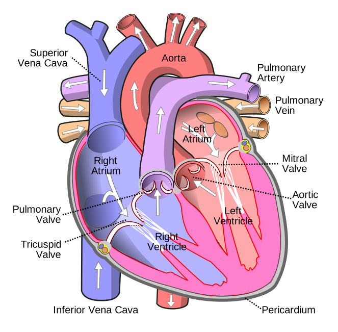
| Look for | How to recognise it |
|---|---|
| The two cavae | The vertical vessel on the left of the page, entering the right atrium from above (superior) and below (inferior). |
| The aorta | The arch over the top, three branches rising to the head and arms. It leaves the left ventricle and curves up and over to the patient’s left, so its root is mid-heart. |
| The pulmonary trunk | In front of the aorta, leaving the right ventricle and splitting into right and left pulmonary arteries; the right one passes behind the aorta. |
| The pulmonary veins | Short stubs entering the left atrium from the sides — usually two each side, four in all. |
| The ventricles | The left has a much thicker wall — the easiest tell on an unlabelled drawing. |
| The apex | The tip, down and to the patient’s left, formed by the left ventricle. Not a stop. |
The Word Bank
The only word bank the course has shown you is on the heart lab worksheet, where you labelled the heart and traced the flow in red and blue. Its terms, sorted by whether each is a stop:
| Term | On the path? | Where it goes |
|---|---|---|
| Superior Vena Cava | Yes | Stop 1 — given on the quiz |
| Inferior Vena Cava | Yes | Stop 1 — given on the quiz |
| Right Atrium | Yes | Stop 2 |
| Tricuspid Valve | Yes | Stop 3 |
| Right Ventricle | Yes | Stop 4 |
| Right Pulmonary Artery | Yes | Stop 6, to the right lung |
| Left Pulmonary Artery | Yes | Stop 6, to the left lung |
| Right Pulmonary Vein | Yes | Stop 8, from the right lung |
| Left Pulmonary Veins | Yes | Stop 8, from the left lung |
| Left Atrium | Yes | Stop 9 |
| Mitral Valve | Yes | Stop 10 |
| Left Ventricle | Yes | Stop 11 |
| Aorta | Yes | Stop 13 |
| Apex | No | A landmark — the tip of the left ventricle. Labelled on the diagram, never filled into a flow blank. |
The documents disagree on ten blanks or eleven (see the endnote); you cannot predict the subset, so learn the whole chain. Part 2 drills every version.
One Name, Several Labels
A blank that wants “mitral” may sit beside a diagram that says “bicuspid”. A change of label is not a change of answer.
| Course name | Also called | Why |
|---|---|---|
| Tricuspid valve | Right AV (atrioventricular) valve | Right atrium to right ventricle; three cusps. |
| Mitral valve | Bicuspid valve · left AV valve | Two cusps, like a bishop’s mitre. Mitral and bicuspid are the same valve. |
| Pulmonic valve | Pulmonary (semilunar) valve | The deck says pulmonic, its valve figure pulmonary. Both are right. |
| Aortic valve | Aortic semilunar valve | Half-moon cusps, like the pulmonic. |
| Pulmonary arteries | Pulmonary trunk, then right & left pulmonary arteries | A blank may want the trunk, the singular or the plural. |
| Vena cava | Superior & inferior · plural venae cavae | Two vessels, one destination — the right atrium. |
The atrioventricular-and-semilunar terminology is standard anatomy rather than something the cardiac deck spells out; it is how the four valves sort into the two heart sounds in Part 3.
The Quiz, Rehearsed
The quiz as the blueprint describes it: venae cavae given, the rest blank, a word bank with Apex left in to reject. Type into each blank, or click a blank and then a word.
Three versions, because the documents disagree on the count. Eleven is right atrium to aorta with all four valves (the peripheral-vascular prep sheet’s count). Thirteen adds the lungs and the body. Nine drops the two semilunar valves, as the worksheet’s bank does. When you can do all three with the bank hidden, you are ready.
Two Kinds of Valve
The four valves come in two pairs, and the pairing is the key to the heart sounds. Each lets blood through one way only — the Think Conceptually handout’s “Normal Function of the Heart Valves (open and closing)”. (Standard physiology: valves have no muscle; they open and shut passively, on pressure.)
| Pair | Valves | Sits between | Shuts when… and makes |
|---|---|---|---|
| Atrioventricular (AV) | Tricuspid (right) Mitral (left) | Each atrium and the ventricle below it | The ventricles start to squeeze — start of systole. Together they make S1, “lub”. |
| Semilunar | Pulmonic (right) Aortic (left) | Each ventricle and its great artery — pulmonary trunk, aorta | The ventricles stop squeezing and relax — start of diastole. Together they make S2, “dub”. |
(Standard anatomy, not in the deck: the AV valves are held by chordae tendineae on papillary muscles, which stop the flaps blowing back; semilunar cusps simply fill and seal.)
Lub and Dub
The deck: systole is “myocardial contraction”, diastole the “filling phase”; Jeopardy: the phase “related to relaxing of the heart muscle” is diastole. The two sounds are the borders between the phases.
| Sound | Valves closing | When | Loudest at |
|---|---|---|---|
| S1 “lub” | Mitral and tricuspid — the AV valves | Start of systole | The apex — tricuspid and mitral areas |
| S2 “dub” | Aortic and pulmonic — the semilunar valves | Start of diastole — “marking the end of systole” | The base — aortic and pulmonic areas |
Drag sideways to see the whole figure →
The clips are played two octaves higher than life so a phone or laptop speaker can carry them; with headphones, open “Original pitch” under each player to hear the true sound a stethoscope gives.
Original pitch, for headphones
Where Each Valve Is Heard — APETM
The largest block on the Exam 4 blueprint: 8 questions with skeletal figures and locating heart sounds. The five listening posts, top to bottom, spell APETM.
Heart lab prep sheet: “APETM designation… understand the landmarks, where S1S2 heard loudest and why (will be tested).”
Exam 4 blueprint: “Must be able to name and document the locations (on a skeletal figure) to auscultate each of the heart sounds. (APETM, full name, and where)”; “Identify the locations of prominent S1 and S2 on the same skeletal figure”; “Auscultating apical pulse and location on a skeletal figure”.
So for each site: the letter, the full name, the intercostal space and border, and which sound is louder there.
| Full name | Where | Louder | Why | |
|---|---|---|---|---|
| A | Aortic | 2nd ICS, right sternal border — the only site on the patient’s right | S2 > S1 | Base. S2 is the aortic and pulmonic valves closing, and the sound carries up, with the flow, into the great arteries. |
| P | Pulmonic | 2nd ICS, left sternal border | S2 > S1 | Base. Same reason, on the pulmonary trunk’s side. |
| E | Erb’s point | 3rd ICS, left sternal border | S1 = S2 | Halfway between base and apex — the deck’s “point at which valves are usually equally audible”. |
| T | Tricuspid | 4th ICS, lower left sternal border | S1 > S2 | Apex. S1 is the tricuspid and mitral closing, and the sound carries down, with the flow, into the ventricles. |
| M | Mitral | 5th ICS, left midclavicular line — the apex, the PMI, the apical pulse | S1 > S2 | Apex. Same reason, at the tip of the left ventricle. |
The lab manual words the tricuspid row “S1 > or = S2”; use the deck’s five lines (see the endnote).
This works like the skeletal figure on Exam 4. Start hides every letter and label and names a site — click where you would put your stethoscope. Then say its intercostal space and which sound is louder there before you look.
Drag sideways to see the whole figure →
Lab manual: normally 1–2 cm and brief, often not palpable in adults, displaced laterally and left in late pregnancy. Slide-16 notes say “4th–5th ICS”; answer fifth, though “4th–5th” is not wrong (endnote).
The technique, in the deck’s order
- Rate and rhythm first, in one area — “usually apex or Erb’s point”. Regular: count 30 seconds and multiply by 2. Irregular: count a full minute.
- Then each area: S1, S2 and any extra sounds.
- Erb’s point is the alternative apical-pulse site — “Often easier than Apex for large breasted women.”
- Pulse deficit = apical − radial (Jeopardy), for an irregular pulse: 96 at the apex, 80 at the wrist → 16 beats too weak to make a pulse. (Standard: two nurses, same full minute.)
- Draping: displace breast tissue laterally; expose only the part of the chest being assessed.
Bell or Diaphragm
Equipment: a stethoscope and a watch with a second hand. Lab manual: “Listen with bell (light pressure) and diaphragm (heavy pressure) of stethoscope at all sites.”
| Side | How to hold it | Best for |
|---|---|---|
| Diaphragm (the wide, flat side) | Firm pressure | High-pitched sounds: S1 and S2, and most murmurs |
| Bell (the small, cupped side) | Light pressure — just enough to seal | Low-pitched sounds: S3 and S4, the rumble of mitral stenosis, and bruits over an artery |
Finding the Spaces by the Ribs
On a drawing, the only thing you can count is ribs, so learn each site as a rib address.
Rib addresses: aortic and pulmonic between ribs 2 and 3, Erb’s 3–4, tricuspid 4–5, mitral 5–6 at the midclavicular line.
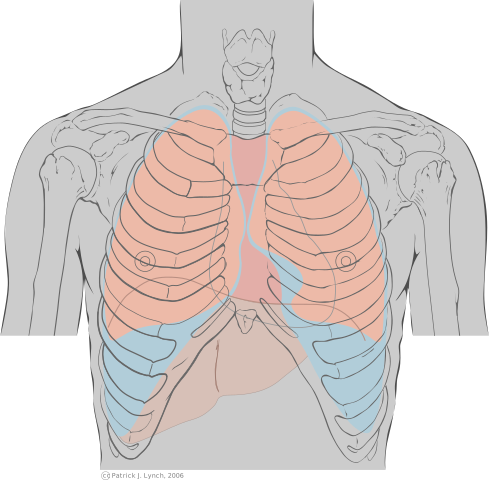
Press Start. The unmarked copy has no dots: count down from the sternal angle and click where each site is. Some questions ask by name, some by rib address, some by which sound is louder.
Where the Valve Is, and Where You Listen
Two maps of the same four valves. Anatomy: all four bunched behind the sternum, from the pulmonic (highest) to the tricuspid (lowest), where their sounds overlap (standard anatomy). Auscultation — APETM — listens downstream instead.
Switch between the two maps, then show both to see how each valve’s sound travels to its post.
Drag sideways to see the whole figure →
| Valve | Where it sits | Where you listen | Why there |
|---|---|---|---|
| Aortic | Behind the left half of the sternum, about the 3rd ICS | 2nd right ICS, RSB | The aorta rises up and to the right before it arches — so a left-sided valve is heard on the right. |
| Pulmonic | At the left 3rd costal cartilage — highest | 2nd left ICS, LSB | The pulmonary trunk runs up and to the left. |
| Tricuspid | Behind the right half, about the 4th ICS — lowest | 4th left ICS, LLSB | Into the right ventricle, behind the lower sternum. |
| Mitral | Behind the left half, about the 4th costal cartilage | 5th left ICS, MCL — the apex | Into the left ventricle, which points down and out. |
| Erb’s point | No valve of its own | 3rd left ICS, LSB | Between base and apex: S1 and S2 about equal. |
Extra Sounds
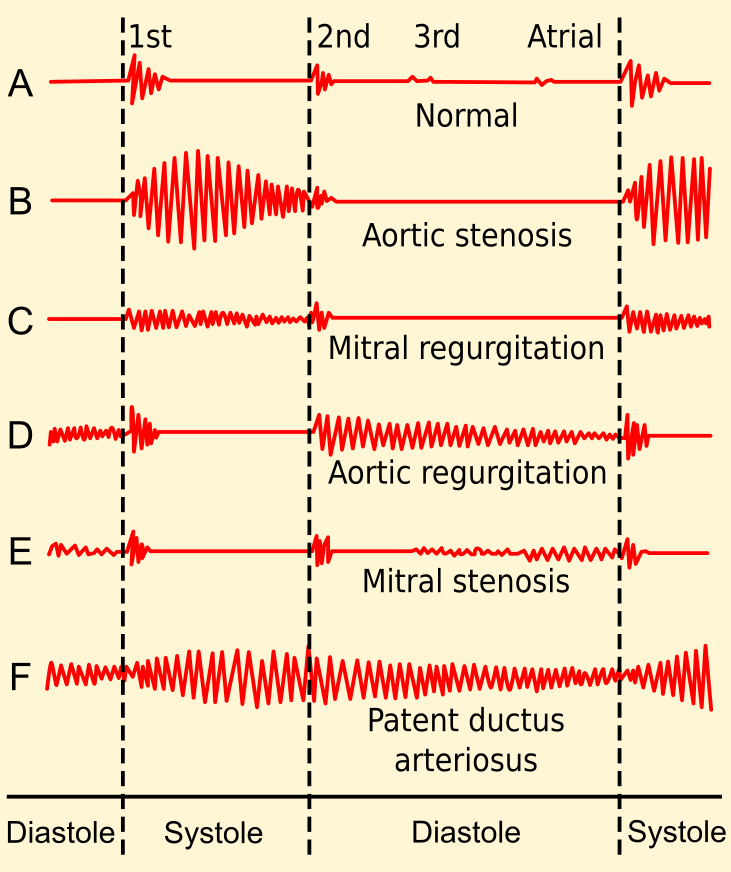
Anything beyond two syllables is an extra sound; the sound strip under Lub and Dub marks where each falls.
S3 and S4 — the gallops
Both are quiet, low thuds in diastole from blood hitting a ventricle that cannot take it. The deck: S3 is “rarely normal in older adults”; S4 comes “late in diastole” from “abnormal resistance to ventricular filling”.
The deck lists children among those in whom S4 can be normal; standard teaching says it is S3 that is normal in children (see the endnote).
| S3 | S4 | |
|---|---|---|
| Name | Ventricular gallop | Atrial gallop (slide 3) — but see the trap below |
| Timing | Early diastole, immediately after S2 | Late diastole, immediately before S1 |
| Cadence | Ken-tuc-ky — lub, dub, ta | Ten-nes-see — ta, lub, dub |
| Mechanism | Rapid ventricular filling into an overfull ventricle | Resistance to ventricular filling — a stiff ventricle |
| Can be normal in | Children, late pregnancy, high cardiac output | Athletes, elderly; the deck adds children (see the note above) — but “usually abnormal” |
| Worrying in | Older adults — heart failure (CHF), fluid overload | CAD, hypertension, MI, aortic or pulmonic stenosis |
| How to hear it | Patient in left lateral position (deck) | Deck silent; standard teaching: both gallops are low-pitched, so the bell, lightly, at the apex. |
Original pitch, for headphones
Original pitch, for headphones
Murmurs
A “blowing or swooshing sound”, “audible when blood volume [is] increased in [the] heart or blood flow is impeded or altered”, from “cardiovascular disease or circulatory problems”. Systolic: just after S1. Diastolic: just after S2. The lab manual: graded 1 to 6; “a diastolic murmur or murmur > grade 3/6 is never innocent”; abnormal aortic murmurs are heard best at Erb’s point.
Original pitch, for headphones
Original pitch, for headphones
The rest of slide 21
| Sound | What the deck says |
|---|---|
| Split sounds | “Normal variation (valves not closing exact same time)” — one sound becomes two. Lab manual: normal split S2 at the pulmonic area, on inspiration; a split S1 at the lower left sternal border possible. |
| Systolic ejection click | “Sudden distention of the great arteries” — early systole, as the semilunar valves open. |
| Opening snap | “Associated with mitral stenosis” — a stiff mitral valve snapping open in early diastole, the exception to “opening is silent”. |
| Pericardial friction rub | Listed without comment — see the next section. |
Original pitch, for headphones
The handout’s sound list adds muffled; in standard teaching that suggests fluid around the heart (pericardial effusion).
Pericardial or Pleural Rub?
Both are a scratchy, grating sound of two membranes rubbing; the difference is what moves them.
- A pleural rub stops — the visceral and parietal pleura rubbing (Jeopardy’s wording), which move only when the lungs move.
- A pericardial rub keeps going, because the heart keeps beating.
| Pleural rub | Pericardial rub | |
|---|---|---|
| Layers rubbing | Visceral and parietal pleura — around the lungs | Layers of the pericardium — the sac around the heart |
| Keeps time with | Breathing | The heartbeat |
| Client holds breath | Stops | Continues |
| Pain it goes with | The deck’s chest-pain list: “Pulmonary disease / Pleural pain” | The same list: “Pericardial pain” |
Standard teaching: ask for only a brief breath-hold; a pericardial rub is often loudest at the lower left sternal border with the client sitting up and leaning forward.
Bruits and Thrills
Murmur, bruit, thrill: one event found three ways — turbulent flow through a narrowed or damaged opening. Where you find it, and whether you hear or feel it, names it. The Exam IV review: a bruit is “a swishing sound… in the carotid artery or any other artery”.
| Where | Heard or felt | What it means | What you do next | |
|---|---|---|---|---|
| Murmur | Over the heart | Heard — blowing | Turbulence through a valve or chamber | Note timing, site and grade; report a new one |
| Bruit | Over an artery — carotid, abdominal aorta, femoral | Heard, with the bell — “swishing” | Narrowing or disrupted flow | Report — a sign of arterial disease (at the neck, possibly an enlarged thyroid) |
| Thrill | Heart or artery | Felt — a fine vibration | Turbulence strong enough to palpate | Listen there: a murmur over the heart, a bruit over a carotid |
| Pericardial rub | Over the heart | Heard — scratchy | Inflamed pericardium, not flow | Breath-hold — it keeps going |
Original pitch, for headphones
The recorded lecture: “we utilize the bell of our stethoscope to listen for bruits. And that’s a swishing sound.” Jeopardy: a bruit “can indicate a narrowing or disruption of blood flow.” HEENT deck: an enlarged thyroid can disturb carotid flow, so auscultate for a bruit. Cardiac deck notes: carotids one at a time, so the brain’s supply is not cut off.
Standard content: feel a thrill with the palm or base of the fingers; it first appears at murmur grade 4. Lab manual: a bruit can be normal in children and high-output states.
The One Rule: Look One Stop Backwards
The Exam 4 blueprint’s version of the heart lab’s “what happens with abnormalities” adds the clue: “Important to understand blood flow and internal structures to answer these types of questions.” You work the symptoms out from the fourteen stops. Blood is a one-way queue: when a chamber or valve fails, blood waits in whatever comes before it, and whatever comes after it goes short — a backward half (congestion) and a forward half (not enough arriving).
Drag sideways to see the whole figure →
| What fails | Backs up into (look back) | Goes short (look forward) |
|---|---|---|
| Tricuspid valve · 3 | Right atrium (2), the superior and inferior vena cava (1), the body. The prior-year review asks exactly this. | Less reaches the right ventricle and the lungs. |
| Right ventricle · 4 | Right atrium, venae cavae, body — neck veins, legs, liver. | Less reaches the lungs, so in the end less reaches the body. |
| High pressure in the pulmonary artery · 6 | The right ventricle pushes harder, tires and fails; then the cavae back up. | Less blood gets through the lungs. |
| Mitral valve · 10 | Left atrium (9), pulmonary veins (8), lungs (7). | Less reaches the left ventricle and the body. |
| Left ventricle · 11 | Left atrium, pulmonary veins, the lungs. | Decreased cardiac output to the whole body. |
Left- and right-sided failure as named syndromes, and the backward/forward framing, are standard nursing content (Taylor Ch 40; ATI Cardiovascular); this note covers every left-versus-right table in this part.
Left Side Fails — the Lungs Fill
When the left ventricle cannot empty — scarred by an MI, or worn by years of high blood pressure — blood waits in the left atrium, pulmonary veins and lungs, and fluid is pushed into the air spaces: every backward sign is wet lungs.
| Finding | Why it happens — and where the course shows it |
|---|---|
| Dyspnea | Fluid in the air spaces gets in the way of gas exchange. “Difficulty breathing” is on the deck’s bolded history list. |
| Orthopnea | Breathless lying flat, better sitting up — lying down returns fluid from the legs and spreads the lung fluid; sitting up lets it settle. The deck’s question: “How many pillows do you use for sleep?” One pillow to three is the lungs filling. |
| Crackles | Air bubbling through fluid, heard first at the bases. HB (anterior-wall MI on a background of hypertension) has “bibasilar crackles”. The Think Conceptually handout: “Auscultate lung sounds — (pulmonary edema associated with cardiac dysfunction)”. |
| Cough | The airways clearing the fluid; on the deck’s past-history slide beside shortness of breath and orthopnea. |
| Fast, shallow breathing · low SpO2 | Less oxygen crosses at stop 7. HB’s respirations are 28/min, shallow. |
Standard nursing content: the mechanisms in the “why” column; the cough of pulmonary edema can bring up frothy, pink-tinged sputum; some patients wake suddenly at night short of breath (paroxysmal nocturnal dyspnea).
The forward half: the left ventricle is the only pump that feeds the body, so its failure is decreased cardiac output — see Not Enough Forward, below.
Right Side Fails — the Body Fills
When the right ventricle cannot keep up, blood waits in the right atrium, the venae cavae and every vein that empties into them — neck above; liver, gut and legs below. The lungs are downstream, so they can stay clear.
| Finding | Why it happens — and where the course shows it |
|---|---|
| Jugular vein distention (JVD) | The jugulars drain straight into the superior vena cava, so the backup shows there first. Jeopardy: “Bulging of this vein can indicate right sided heart failure”. Handout: “Assess for jugular vein distention (venous congestion from heart failure)”. The deck lists it under inspection. |
| Peripheral, dependent edema | Fluid pushed into the tissues where gravity takes it — feet and ankles up and about, the sacrum in bed — usually both sides. On the deck’s history list and cues. |
| Weight gain | About a kilogram a litre, so the scale shows it before the ankles. Deck cue “Weight Gain”; handout “Assess Daily weights — fluid status”. |
| Enlarged liver, swollen abdomen | The liver drains into the inferior vena cava, so it swells (hepatomegaly) and fluid can collect in the abdomen. HB’s form checks for it — “negative hepatomegaly”. |
Standard nursing content: abdominal congestion can cause loss of appetite and nausea; the abdominal fluid is ascites. The lab manual gives the normal finding as no neck vein distention, jugular venous pressure under 3 cm with the patient at 45°.
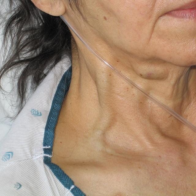
“What assessment finding is associated with tricuspid valve dysfunction (blood backing up from the dysfunctional valve)?” It “would back-up blood flow in the Superior/Inferior Vena Cava”: JVD and peripheral edema.
“Which ventricle’s workload will increase if there is high pressure in the pulmonary artery?” The right — it empties into that artery, so the pressure is its afterload. The handout: “high pressure — heart has to work harder — failure”.
Standard nursing content: chronic lung disease, raising pulmonary artery pressure, is the other classic cause — one reason the deck’s history slide asks about “Lung disease”.
Why Heart Patients Get Up at Night
Nocturia is getting up during the night to urinate — Jeopardy’s “A nurse charts this term when a patient reports having night time urination.” Three steps:
1. By day, the fluid sits in the legs as dependent edema, out of the circulation — the weak heart is not pumping it and the kidneys are not seeing it.
2. At night, lying flat brings it back. The fluid moves from the tissues into the veins, the resting heart pumps it on, and more blood reaches the kidneys.
3. More blood through the kidneys makes more urine overnight — so the patient wakes to urinate, and the ankles are smaller by morning.
The prior-year review: “Nocturia (frequent night urination) and Edema are symptoms of which Heart Condition?” — heart failure. The deck pairs them on its bolded history list and asks “Do you get up during the night to urinate?” right after the pillows question: orthopnea is the lungs’ answer to lying down, nocturia the kidneys’.
The mechanism is standard physiology (Taylor Ch 40). Nocturia has other causes — prostate, diabetes, an evening diuretic, bedtime fluid — so look for it paired with edema.
Not Enough Forward: Decreased Cardiac Output
Cardiac output: Jeopardy’s “how much blood the left ventricle pumps out in a minute”, “a reflection of blood flowing around the body, especially to the brain and other vital organs.”
Standard: resting output is roughly 4–8 litres a minute; the compensating rise in rate costs the heart oxygen.
The short answer is decreased tissue perfusion — the prior-year review’s “How does it affect tissue perfusion?”
| Where | What you find | Source |
|---|---|---|
| Circulation | Blood pressure falls; heart rate rises to compensate; pulses weak (+1). | Jeopardy: BP is “highly reflective of cardiac output”. HB: +1 peripheral pulses. |
| Brain | Dizziness, restlessness, confusion, a change in level of consciousness. | Handout: “Monitor for changes in level of consciousness (blood getting to brain?)”. Dizziness is on the deck’s history list. |
| Kidneys | Less urine; BUN and creatinine rise. | Handout: “BUN, Creat — elevate if impaired blood flow to Kidney as in Heart Failure”; “Monitor Intake and Output”. |
| Skin & extremities | Cool, pale skin, slow capillary refill, weak peripheral pulses. | HB: skin “pale/ashen”, “poor capillary refill”, skin cool. |
| Muscles | Fatigue, tiring easily, unable to keep up with daily activities. | Deck history list: “Fatigue/ tire easily”; lifestyle slide: “Can you perform daily activities?” |
| The heart itself | Chest pain — the heart muscle is short of oxygen too. | Deck: cardiac pain is “cells deprived of O2”. |
Standard: on a nitroglycerin drip, like HB, BP is also what the drug can drop.
| Watching for | Interventions (handout wording) |
|---|---|
| Forward failure — output | Monitor vital signs (temp, pulse rate and rhythm, respirations, BP) · EKG · changes in level of consciousness · labs (Hgb, K, Mg, creat) |
| Backward failure — lungs | Auscultate lung sounds · oxygen as indicated · position for improved ventilation — “increased lung expansion, improve venous return” |
| Backward failure — body | Jugular vein distention · peripheral edema · intake and output · daily weights |
| The heart itself | Heart sounds (extra sounds, murmur, muffled, rub) · assess chest pain, treat promptly · bedrest if acute MI or CHF exacerbation — “decrease workload of heart” — then progressive activity |
Left-Sided Versus Right-Sided, Side by Side
Sort a stem’s findings into the columns; whichever fills is the failing side. Both? The left probably went first.
| Left side fails | Right side fails | |
|---|---|---|
| The failing pump | Left ventricle (11), or the mitral valve (10) before it | Right ventricle (4), or the tricuspid valve (3) before it |
| Backs up into | Pulmonary veins → lungs | Venae cavae → body |
| Lung sounds | Crackles, at the bases first | Usually clear — unless the left side is failing too |
| Breathing | Dyspnea, orthopnea, cough, fast shallow respirations, low SpO2 | Not the main complaint |
| Neck veins | Flat, at first | JVD |
| Edema & weight | Not the first sign | Dependent edema, both legs or the sacrum · weight gain |
| Abdomen | — | Enlarged liver, swollen abdomen |
| Forward shortfall | Decreased cardiac output to the body: falling BP, fatigue, dizziness, LOC changes, cool pale skin, less urine | Less blood to the lungs, and so in the end to the left heart and the body |
| The deck’s question that finds it | “How many pillows do you use for sleep?” | “Edema or swelling” |
| Either side | “Do you get up during the night to urinate?” — nocturia points to heart failure, not to one side | |
| Stem words that point here | lungs, crackles, short of breath, pillows, sits up to breathe, cough, mitral | neck veins, JVD, ankles, swelling, weight up, liver, tricuspid, cavae, pulmonary artery pressure |
| Usual cause | MI, long-standing high blood pressure (HB has both) | Left-sided failure; lung disease; high pulmonary artery pressure |
- Left, backward: bibasilar crackles; respirations 28, shallow.
- Left, forward: skin pale/ashen, cool, poor capillary refill, +1 peripheral pulses; heart rate 115.
- Right: “Jugular Vein Distention Note[d]” — but negative hepatomegaly, no edema recorded.
- Heart sounds: a positive S3.
HB’s case gives both “poor capillary refill” and “< 3 seconds”, so build no answer on his refill time. That an anterior-wall MI involves the left ventricle (left anterior descending artery) is standard anatomy, not stated in the case.
Traps
The Exam 4 Blueprint, Mapped
The quiz is one sequence; Exam 4 is the whole unit. The Exam 4 study guide carries this guide’s Parts 1 to 4 and adds the cardiac assessment, peripheral vascular and rest and sleep parts, so the blueprint is mapped line by line there.
One line from it that this guide’s practice quiz asks about: the cardiac deck’s only red line, “Consider all chest pain to be cardiac until cardiac issue is ruled out” — gather data while treating with O2 and nitroglycerin.
The Week’s Terms
Both preparation sheets’ “Define Terms” lists, in the decks’ words where they give one.
| Term | In one line |
|---|---|
| Apex of the heart | The bottom — tip of the left ventricle, 5th left ICS, midclavicular line. |
| Base of the heart | The top, where the great vessels attach; S2 louder here. |
| Dysrhythmia | “Abnormality of rhythm” (deck); Jeopardy’s arrhythmia. |
| Systole | “Myocardial contraction” — ejection. Starts with S1. |
| Diastole | “Filling phase.” Starts with S2. |
| Murmur | A blowing or swooshing sound (Part 3). |
| Preload | “Degree of stretch on the ventricle.” |
| Afterload | “Pressure the ventricle has to pump against.” |
| Palpitation | The client’s awareness of irregular, skipped or pounding beats — subjective. |
| Jugular vein distention (JVD) | Bulging neck veins — backup from the right side. |
| Point of maximal impulse (PMI) | Where the heartbeat is best felt on the chest wall (Part 3). |
| Edema | Swelling from fluid in the tissues, graded +1 to +4 by pit depth — venous trouble or heart failure. |
| Atherosclerosis | “Narrowing from plaque” (PV deck) — the commonest cause of PAD and coronary disease. |
| Arteriosclerosis | “Thickening/hardening of arteries” (PV deck) — calcification with age; raises systolic BP. |
| Thrombus | A clot that forms and stays in a vessel, often where the lining is injured. |
| Embolism | A clot or other mass that has broken loose and travelled, blocking a vessel downstream. |
| Deep vein thrombosis (DVT) | A thrombus in a deep vein — risk of pulmonary embolism. Warm, swollen, tender limb. |
| Varicosities | “Distended, protruding veins that appear darkened and tortuous” (tidbits) — weakened walls and valves. |
| Angina | Chest pain or pressure from too little oxygen to the heart muscle — chest, arm, jaw; stable or unstable. |
| Bruit | A swishing sound over an artery (Part 3). |
Thrombus and embolism are listed but not defined in the decks; those two lines are standard definitions.
HEENT — Key Points
Lectured in the quiz week but not on the Exam 4 blueprint (the syllabus lists it with Exam 4), so key points only.
| Area | What to know |
|---|---|
| Trachea · neck muscles | Midline · turn against resistance, shrug — CN XI. |
| Thyroid | Not palpable; have the patient swallow. Slightly enlarged can be normal in pregnancy. Labs TSH, T3, T4. Enlarged → auscultate for a carotid bruit. |
| Lymph nodes | Finger pads, gentle circles, compare sides; 1 cm nodes can be normal at ages 1–5. |
| Temporal arteritis | Biopsy, sed rate; high-dose steroid; untreated → blindness. |
| Vision | Snellen (“E” chart if no English) · near vision over 40 · confrontation fields, Ishihara colour · PERRLA · sudden loss: emergency referral; gradual is not urgent. |
| Eye muscles · red reflex | Six cardinal fields of gaze, CN III, IV, VI; corneal light reflex, cover test · absent red reflex may mean a lens opacity. |
| Hearing loss | Conductive: blocked on the way in (cerumen, middle-ear fluid) · sensorineural: nerve. Hearing loss makes people more prone to depression (deck); cotton swabs cause impaction. |
| Whisper · otoscope | Out of sight of your mouth, one ear at a time, 18 inches · adult pinna up and back; eardrum transparent, intact, not bulging. |
| Otalgia · balance | Ear pain: most commonly otitis media · Romberg. |
| Nose · sinuses | Septum midline · frontal, ethmoid, maxillary: palpate, percuss · abrupt anosmia → MRI (possible cancer). |
| Mouth · throat | White patches: Nystatin early · oral cancer: tobacco, HPV, alcohol, alcohol-based mouthwash · uvula midline, gag reflex, swallowing. |
| Emergencies | ABCs: epistaxis is an airway problem first; in anaphylaxis the epiglottis may block the airway. |
| Hypothyroidism — slowed down | Hyperthyroidism — sped up |
|---|---|
| Fatigue | Fatigue |
| Cold intolerance | Heat intolerance, diaphoresis |
| Weight gain or difficulty losing; anorexia | Weight loss |
| Dry skin, brittle hair | Fine, limp hair |
| Menstrual irregularity, decreased libido | Anxiety, palpitations, rapid pulse, muscle weakness |
| Rinne | Weber | |
|---|---|---|
| Where | Stem on the mastoid process; when the client no longer hears it, move the fork beside the ear | Stem on the middle of the skull (midline parietal bone) |
| Compares | Air against bone, in one ear | One ear against the other, by bone |
| Normal | Air conduction about twice as long as bone | Heard equally in both ears |
Drag sideways to see the whole figure →
Whisper distance and the Rinne ratio follow the lecture; the handout’s figures are in the endnote.
The case (LT): BP 186/108 after ten years of hypertension, out of medication; soft bilateral carotid bruit — a stroke risk; chews tobacco; alcohol-based mouthwash; cerumen blocking both ears.
Define list (standard definitions): amblyopia, lazy eye · myopia, nearsighted · glaucoma, raised eye pressure damaging the optic nerve · cataract, clouded lens · macular degeneration, central vision loss · retinopathy, retinal-vessel damage · dysphagia, difficulty swallowing · halitosis, bad breath · tinnitus, ringing · vertigo, the room spinning · epistaxis, nosebleed · conjunctivitis, pink eye · Eustachian tube, middle ear to nasopharynx · vestibular, inner-ear balance · tympanic membrane, eardrum · maxilla / mandible, upper / lower jaw.
The marked items, one more time
| Item | Answer |
|---|---|
| The quiz — “Memorize/understand the blood flow through the heart” | |
| The path | Venae cavae, right atrium, tricuspid, right ventricle, pulmonic, pulmonary arteries, lungs · pulmonary veins, left atrium, mitral, left ventricle, aortic, aorta, body |
| The four valves | Tricuspid, pulmonic, mitral, aortic — Try Pulling My Aorta |
| Artery or vein | Named by direction, not oxygen: pulmonary arteries carry deoxygenated blood |
| Right and left | The patient’s — on a diagram, their right is on your left |
| The format | Word bank, starting at the venae cavae, 10 points; Apex is not a stop |
| Exam 4 — the marked lines in this guide | |
| The block arrows | Diastole → filling. Systole → ejection of blood. S1 (AV valves) starts systole; S2 (semilunar) diastole |
| APETM | A 2nd ICS RSB · P 2nd ICS LSB · E 3rd ICS LSB · T 4th ICS LLSB · M 5th ICS left MCL. “You will be tested on this” |
| Where S1 and S2 are loudest | Base S2 · Erb’s equal · apex S1 — near, not over, the valves; sound radiates with the flow |
| PMI | 5th ICS, midclavicular line = apex = apical pulse = mitral area |
| S3 · S4 | S3 after S2: heart failure, fluid overload · S4 before S1: CAD, HTN, MI, stenosis |
| Pleural or pericardial rub | Hold the breath: pleural stops, pericardial keeps going |
| Carotid | Bell for a bruit (narrowing); enlarged thyroid → listen; palpate one side at a time |
| Left vs right failure | Left → lungs: crackles, dyspnea, orthopnea · right → cavae → body: JVD, dependent edema, weight gain |
| Nocturia | Lying flat returns leg fluid to the kidneys. With edema → heart failure |
| Decreased cardiac output | Decreased tissue perfusion; the priority vital sign is BP |
| Weber · Rinne (highlighted; HEENT, for the final) | Weber: midline skull, equal both ears · Rinne: mastoid then beside the ear, air twice as long as bone |
The night before the quiz: the first five rows above are the whole quiz.
Other things worth knowing
| Item | Answer |
|---|---|
| Two pumps | Same volume every beat; the left is stronger, not busier |
| Thickest wall | Left ventricle — the tell on an unlabelled drawing |
| Other names | Mitral = bicuspid · pulmonic = pulmonary |
| The drill | Eleven with the bank → bank hidden → the nine → Trace it |
| Item | Answer |
|---|---|
| Which sound is S1 | The one after the long pause |
| Coronary arteries | Fill in diastole |
| Apical rate | Regular: 30 s × 2. Irregular: a full minute. Erb’s point is the alternative site |
| Pulse deficit | Apical − radial |
| Rib count | Sternal angle = rib 2; a space is named for the rib above it |
| Bell · diaphragm | Bell, light: S3, S4, bruits · diaphragm, firm: S1, S2 |
| Murmurs | Systolic after S1, diastolic after S2; diastolic or above 3/6 never innocent |
| Other extra sounds | Split S2: normal, pulmonic area, inspiration · opening snap: mitral stenosis |
| Felt a thrill | Listen at that spot |
| Item | Answer |
|---|---|
| The one rule | Backs up one stop back; mitral → lungs, tricuspid → cavae |
| Orthopnea | “How many pillows do you use for sleep?” |
| Commonest cause of right-sided failure | Left-sided failure |
| Cardiac output | SV × HR; preload = stretch, afterload = pressure pumped against |
| The deck’s three problems | Impaired cardiac output, impaired tissue perfusion (forward) · excess fluid volume (backward) |
| Chest pain | Cardiac until ruled out (deck, in red) |
| Sclerosis · clots | Arterio hardening, athero plaque · thrombus stays, embolus travels |
| HEENT | Adult otoscope pinna up and back · no red reflex → lens opacity |
Traps, one line each
| Trap | Correct answer |
|---|---|
| No semilunar valves in the bank | Not off the quiz — learn all four |
| S1 as the filling sound | S1 starts systole; filling is the silence after S2 |
| Aortic heard on the left | The only site on the patient’s right |
| “Located” vs “heard” | Located = behind the sternum; heard = the listening post |
| S4 as “ventricular gallop” | S4 is the atrial gallop |
| S4 “normal in children” | It is S3 that is normal in children |
| Cough to sort a rub | Cough is for crackles and rhonchi |
| “Hear a thrill” | A thrill is felt, never heard |
| High pulmonary artery pressure | Overworks the right ventricle |
| Crackles in a cardiac stem | Left-sided failure before pneumonia |
| Edema always means the heart | One warm, tender leg = DVT; both plus JVD = heart |
| BP still high, so output is fine | Watch BP because it will show the fall |
| Shiny, hairless legs after an MI | Months of arterial insufficiency, not the MI |
| Fatigue and the thyroid | On both lists; use temperature and weight |
Read the recap
Recap: The Path, Drawn
The whole quiz is one chain, and the quiz hands you the first link, the venae cavae. Say the rest as two sentences. Venae cavae, right atrium, tricuspid, right ventricle, pulmonic, pulmonary arteries, lungs. Pulmonary veins, left atrium, mitral, left ventricle, aortic, aorta, body. The valves in order are tricuspid, pulmonic, mitral, aortic: Try Pulling My Aorta. Remember three things. An artery leaves the heart, so the pulmonary arteries carry blue blood. Right and left are the patient’s, so the right heart is on your left. And Apex is a landmark, not a stop. The worksheet’s bank leaves out the pulmonic and aortic valves; learn them anyway.
Recap: Drill It
Part 2 is practice. Do the eleven blanks with the bank showing, then hide the bank and do it again. Then do the nine, which jumps from each ventricle straight to its artery. When every version comes out clean with the bank hidden, you are ready.
Recap: Valves, Sounds and the Cardiac Cycle
A heart sound is a pair of valves shutting. S1, the lub, is the mitral and tricuspid closing as systole starts; S2, the dub, is the aortic and pulmonic closing as diastole starts. Diastole means filling, systole means ejection. Then the five listening posts, top to bottom: aortic and pulmonic in the second space either side of the sternum, Erb’s point in the third, tricuspid in the fourth at the lower left sternal border, and mitral in the fifth at the midclavicular line, which is also the apex and the PMI. S2 is louder at the base, S1 at the apex, and they are equal at Erb’s point, because the posts sit near the valves, not over them, and sound follows the flow. S3 comes after S2, like Kentucky, and means heart failure; S4 comes before S1, like Tennessee, and means a stiff ventricle. A swoosh over the heart is a murmur, over an artery a bruit, and a thrill is felt, never heard. For a rub, ask the client to hold their breath.
Recap: When the Flow Backs Up
Work the symptoms out from the path: a failure backs blood up into the stop before it, and leaves the stop after it short. Left goes to the lungs: crackles at the bases, shortness of breath, and orthopnea, found by asking about pillows. Right goes to the rest of the body: distended neck veins, swelling in both legs, weight gain and an enlarged liver. A failing tricuspid backs up into the venae cavae, and high pulmonary artery pressure overworks the right ventricle. Nocturia happens because lying flat returns the leg fluid to the kidneys. When output falls, every organ is short of blood, and blood pressure is the vital sign to watch.
Recap: Beyond the Quiz
The Exam 4 guide maps the rest of the blueprint. Keep the deck’s red line: consider all chest pain cardiac until ruled out. Arteriosclerosis is hardening and atherosclerosis is plaque; a thrombus stays and an embolus travels. For HEENT, Weber is the fork on the middle of the skull, equal in both ears; Rinne is the mastoid and then beside the ear, with air lasting about twice as long as bone. An enlarged thyroid means listening for a carotid bruit.
Sources. The course’s own material, which always takes precedence: the Quiz 3 and Exam 4 blueprints; the heart lab, peripheral-vascular and HEENT-week preparation sheets; the APETM lab activity; the cardiac lab deck and its speaker notes; the heart lab worksheets (the blood-flow diagram and word bank, and the APETM chest figures); the “path of blood through the heart” handout; the Cardiovascular System: Think Conceptually handout and the cardiac case summary (HB); the lab manual’s cardiovascular and peripheral vascular tables (facts only; no pages reproduced); the peripheral vascular lecture deck and its recorded lecture, the PVD/PAD handout; the HEENT lecture deck and assessment summary and the head-and-neck case (LT); the syllabus; the Exam 4 Jeopardy review; and a prior year’s Exam 4 review sheet. Chapter references are to Taylor, Fundamentals of Nursing — Ch 40 Oxygenation and Perfusion.
Where the course documents disagree with each other. Each is flagged where it matters rather than smoothed over. The quiz is ten blanks in the blueprint and the HEENT-week plan but eleven on the peripheral-vascular preparation sheet; the guide drills every version. The lab worksheet’s word bank has no pulmonic or aortic valve, and it includes Apex, which is not a stop; the guide drills all four valves anyway. The cardiac deck’s terminology slide calls S3 the ventricular gallop and S4 the atrial gallop, while its extra-sounds slide groups both under “ventricular gallop”; the guide follows the terminology slide. The prior year’s review sheet carries arterial blood gas items that belonged to that year’s Exam 4; this year they were on Exam 3, and they are left out. The whisper test is 18 inches in the deck and two to three feet in the HEENT summary, and the Rinne ratio is “twice” or “two to three times”; the deck is followed. The lab manual words the tricuspid area (lower left sternal border) as “S1 > or = S2”; the deck’s S1 louder there, and S1 = S2 at Erb’s point, are used. The slide-16 notes place the PMI at the “4th–5th ICS” where the slide says fifth; fifth is used. For the priority vital sign when cardiac output falls, the prior year’s review hints at the blood’s oxygen level while Jeopardy answers BP twice; BP is used. HB’s case gives both “poor capillary refill” and “< 3 seconds”, so no answer is built on it. One deck line is treated as a slip and flagged: cardiac slide 3 lists children among those in whom S4 can be normal (standard teaching says that is S3). The peripheral vascular and sleep conflicts are listed in the Exam 4 guide, where that content lives. The syllabus lists HEENT with Exam 4; the Exam 4 blueprint does not — the blueprint is followed, and HEENT is summarised in Part 5.
The illustrations and sounds. Six heart-sound recordings, a carotid bruit and four figures are reproduced under open licences, each credited where it appears: the systolic murmur is EmilyHopeS, CC BY-SA 4.0; the normal heart sounds, S3, S4, diastolic murmur and split S2 are from the University of Michigan Heart Sound & Murmur Library (Judge and Mangrulkar), CC BY-SA 3.0; the carotid bruit is James Heilman, MD, CC BY-SA 3.0; the heart interior is by Wapcaplet, the phonocardiograms by Madhero88 and the jugular vein photograph by Ferencga, all CC BY-SA 3.0, and the rib-cage figure by Põnn after Patrick J. Lynch, CC BY 2.5; all four are from Wikimedia Commons. Audio clips were trimmed and re-encoded, and each is played two octaves higher, compressed and loudness-matched so a phone speaker can carry it, with the true-pitch recording in a fold beneath; the photograph was resized and the rib-cage figure’s original dots removed; each adapted file remains under its original licence. The loop flow chart in Part 1 and every other schematic not credited here are original drawings made for this guide. The decks’ own images, the lab worksheets’ figures and the lab-manual pages are instructor and publisher material and are not reproduced anywhere here.
Grey type, textbook content and mnemonics. Notes in the smaller grey type are either standard nursing content, used sparingly — the atrioventricular and semilunar grouping, the alternative valve names, the physiology behind backward failure and nocturia — or a course document’s figure that conflicts with what you were told, kept small so it does not compete. Where a course document says something, that is what the guide answers with; where the two ever conflict, what you were told in class wins. Try Pulling My Aorta and the other memory sentences are study aids added for this guide, not course material; the facts they carry are the course’s.